. 2022 Jun 7;12(1):238.
doi: 10.1038/s41398-022-02005-w.

Prenatal hypoxia alters the early ontogeny of dopamine neurons

Affiliations

Prenatal hypoxia alters the early ontogeny of dopamine neurons

Anastasia Brandon et al. Transl Psychiatry. .

Abstract

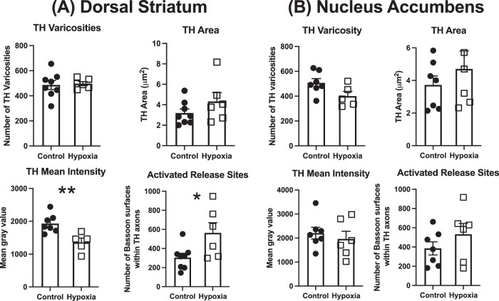
Dopaminergic (DA) dysfunction is a significant feature in the pathophysiology of schizophrenia. Established developmental risk factors for schizophrenia such as maternal immune activation (MIA) or developmental vitamin D (DVD) deficiency, when modelled in animals, reveal the differentiation of early DA neurons in foetal brains is delayed suggesting this may be a convergent aetiological pathway. Here we have assessed the effects of prenatal hypoxia, another well-known developmental risk factor for schizophrenia, on developing DA systems. Pregnant mice were exposed to a hypoxic environment of 10% oxygen for 48 h from embryonic day 10 (E10) to E12. Embryonic brains were collected and the positioning of mesencephalic cells, expression of DA specification and maturation factors were examined along with the expression of factors that may govern the migration of these neurons. We show that prenatal hypoxia results in a decrease in dopaminergic progenitors retards early DA neuron lateral migration and reduces expression of the receptors known to govern this process. A second time-point, postnatal day 10 (P10) was also examined in order to assess whether prenatal hypoxia alters early presynaptic architecture in the developing striatum. We show reduced expression of tyrosine hydroxylase (TH) in the postnatal striatum along with increases in the density of high-probability DA release sites within TH varicosities. These findings add to the emerging literature showing that multiple epidemiologically validated environmental risk factors for schizophrenia may induce early alterations to develop DA systems. This may represent a possible convergent mechanism in the onset of presynaptic DA dysfunction in patients.


Conflict of interest statement

The authors declare no competing interests.

Figures

Fig. 1
Fig. 1. Prenatal hypoxia alters the early ontogeny of DA neurons.
A Dopaminergic (DA) progenitors (Lmx1a+/Sox2+). There was a reduction in DA progenitor number (Lmx1a+/Sox2+); no difference in Lmx1a expression (mean Grey value); a reduction in DA progenitor mediolateral (X) positioning (relative to midline); and an increase in DA progenitor dorsoventral positioning (Y) (from the ventral boundary of the mesencephalic aqueduct) in E12 hypoxic brains. Image shows brain region examined. A anterior, P posterior. (Control, n = 5; Hypoxia, n = 6; data are shown as mean ± SEM; Group differences are indicated *p < 0.05, **p < 0.01, ***P < 0.001). B Postmitotic (Lmx1a+/Sox2-) cells Hypoxia had no effect on the number of postmitotic (Lmx1a+/Sox2−) cells; no effect on Lmx1a expression (mean Gray value); no effect on mediolateral lateral (X), positioning (relative to midline); and no effect on dorsoventral positioning (Y) (from the ventral boundary of the mesencephalic aqueduct) in E12 hypoxic brains. Image shows brain region examined. A anterior, P posterior. (Control, n = 5; Hypoxia, n = 6; data are shown as mean ± SEM). C Differentiated or mature dopaminergic neurons (TH+) Hypoxia had no effect on the number of mature dopaminergic neurons (TH+); no effect on TH expression; however, there was a reduction in mediolateral (X), positioning (relative to midline); but no alteration in dorsoventral positioning (Y) (from the ventral boundary of the mesencephalic aqueduct) in E12 hypoxic brains. There was also a group/section interaction where the rostrocaudal reduction in TH+ neurons seen in controls was absent in hypoxic brains. Image shows brain region examined. A anterior, P posterior. (Control, n = 4, Hypoxia, n = 6; data are shown as mean ± SEM; Group differences are indicated ***P < 0.001).
Fig. 2
Fig. 2. Prenatal hypoxia reduces the expression of receptors governing lateral DA neuron migration of early DA neurons.
Hypoxia reduced the expression of the receptors DAB1 and PTPRZ1. However, hypoxia had no effect on the expression of their respective ligands Reelin and L1CAM. (Control, n = 12, Hypoxia, n = 12; data are shown as mean ± SEM; Group differences are indicated **P < 0.01).
Fig. 3
Fig. 3. Prenatal hypoxia alters DA terminal architecture in the P10 dorsal striatum.
A There was no effect of prenatal hypoxia on TH varicosity number or area covered by TH varicosities in P10 dorsal striatum. Prenatal hypoxia led to a significant reduction in the intensity of TH expression and increased the number of activated release sites. B There was no effect of prenatal hypoxia on TH varicosity number, area covered by TH varicosities, intensity of TH staining or activated release site number in P10 nucleus accumbens. (Control, n = 8, hypoxia, n = 6; data are shown as mean ± SEM; Group differences are indicated by *p < 0.05, **p < 0.01).
Fig. 4
Fig. 4. Summary figure of the effects of prenatal hypoxia on dopamine neuron ontogeny.
A Prenatal hypoxia induces early changes progenitor cell number, progenitor lateral and ventral positioning and a reduction in lateral positioning of more mature TH+ neurons. B This correlates with a postnatal reduction in the intensity of TH expression and a potentially compensatory increase in presynaptic release sites in TH-positive terminals. Importantly these findings are in the dorsal striatum not the nucleus accumbens consistent with the anatomical loci of presynaptic DA abnormalities reported in schizophrenia.

Similar articles

Cited by

References

    1. Sekar A, Bialas AR, de Rivera H, Davis A, Hammond TR, Kamitaki N, et al. Schizophrenia risk from complex variation of complement component 4. Nature. 2016;530:177–83. doi: 10.1038/nature16549. - DOI - PMC - PubMed
    1. Brown AS. Epidemiologic studies of exposure to prenatal infection and risk of schizophrenia and autism. Dev Neurobiol. 2012;72:1272–6. doi: 10.1002/dneu.22024. - DOI - PMC - PubMed
    1. Eyles DW. How do established developmental risk-factors for schizophrenia change the way the brain develops? Transl Psychiatry. 2021;11:158. doi: 10.1038/s41398-021-01273-2. - DOI - PMC - PubMed
    1. Carlsson A. The current status of the dopamine hypothesis of schizophrenia. Neuropsychopharmacology. 1988;1:179–86. doi: 10.1016/0893-133X(88)90012-7. - DOI - PubMed
    1. Girgis RR, Zoghbi AW, Javitt DC, Lieberman JA. The past and future of novel, non-dopamine-2 receptor therapeutics for schizophrenia: a critical and comprehensive review. J Psychiatr Res. 2019;108:57–83. doi: 10.1016/j.jpsychires.2018.07.006. - DOI - PubMed

Publication types