. 2022 Jun 7;13(1):3263.
doi: 10.1038/s41467-022-30614-y.

JMJD3 intrinsically disordered region links the 3D-genome structure to TGFβ-dependent transcription activation

Affiliations

JMJD3 intrinsically disordered region links the 3D-genome structure to TGFβ-dependent transcription activation

Marta Vicioso-Mantis et al. Nat Commun. .

Abstract

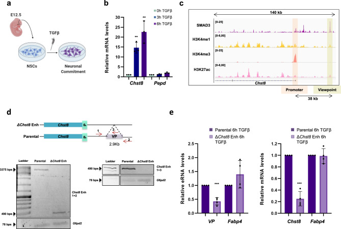
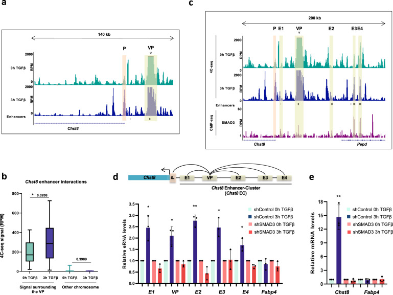
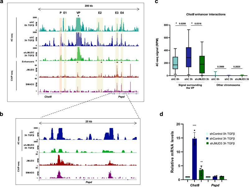
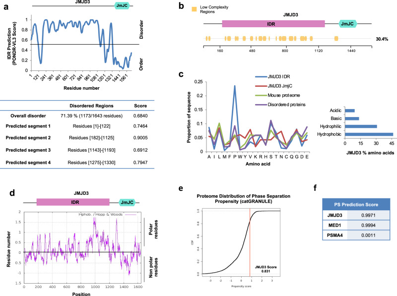
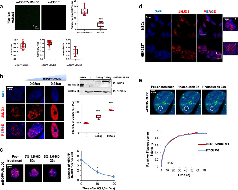
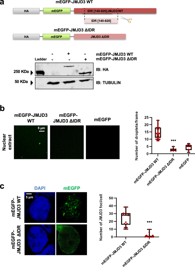
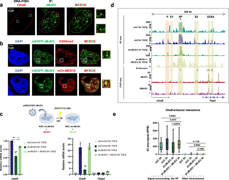
Enhancers are key regulatory elements that govern gene expression programs in response to developmental signals. However, how multiple enhancers arrange in the 3D-space to control the activation of a specific promoter remains unclear. To address this question, we exploited our previously characterized TGFβ-response model, the neural stem cells, focusing on a ~374 kb locus where enhancers abound. Our 4C-seq experiments reveal that the TGFβ pathway drives the assembly of an enhancer-cluster and precise gene activation. We discover that the TGFβ pathway coactivator JMJD3 is essential to maintain these structures. Using live-cell imaging techniques, we demonstrate that an intrinsically disordered region contained in JMJD3 is involved in the formation of phase-separated biomolecular condensates, which are found in the enhancer-cluster. Overall, in this work we uncover novel functions for the coactivator JMJD3, and we shed light on the relationships between the 3D-conformation of the chromatin and the TGFβ-driven response during mammalian neurogenesis.


Conflict of interest statement

The authors declare no competing interests.

Figures

Fig. 1
Fig. 1. The VP is an essential Chst8 enhancer.
a Schematic view of the model used in this study. NSCs were dissected from cerebral cortices of mouse fetal brains (E12.5) and cultured ex vivo (see methods). TGFβ addition leads to neuronal commitment. b NSCs were treated with TGFβ. Total RNA was prepared and the levels of the mRNA of the indicated genes were determined by qPCR. Values were normalized to the housekeeping gene Gapdh and the figure shows values relative to time 0 h. Results are the mean of three biologically independent experiments. Data are presented as mean values +/− SEM. **p < 0.01 (P values were calculated using two-tailed Student’s t test, p = 0.001131677 and p = 0.006143072). Source data are provided as a Source Data file. c UCSC captures showing the chromatin landscape and SMAD3 binding around the Chst8 gene promoter and the Chst8 putative enhancer (VP) in NSCs. Tracks display ChIP-seq in NSCs treated with TGFβ (SMAD3) or untreated NSCs (H3K4me1, H3K27ac, and H3K4me3). Promoter and VP enhancer are shaded in light orange and yellow respectively. d Schematic representation of the CRISPR/Cas9 experimental approach used to delete the Chst8 putative enhancer in NSCs. Two gRNAs flanking the Chst8 enhancer region were used to create the deletion (2.9 kb). Red arrows represent primers to test the deletion. PCRs using Chst8 deletion and G6pd2 pairs of primers are shown at the bottom of the figure in parental and ΔChst8 enh NSC lines. Results are representative of three independent experiments. Source data are provided as a Source Data file. e Parental and ΔChst8 enh cell lines were treated with TGFβ for 6 h. Total RNA was prepared and the levels of eRNA of the VP enhancer (left) or Chst8 mRNA (right) were determined by qPCR. mRNA and eRNA levels of Fapb4 were used as a control. Values were normalized to the Gapdh gene, and figure shows values relative to parental line. Data are presented as mean values +/− SEM. Results are representative of three biological independent experiments. ***p < 0.001 (P values were calculated using two-tailed Student’s t test, p = 2.2027E-05 and p = 1.71676E-08). Source data are provided as a Source Data file.
Fig. 2
Fig. 2. TGFβ drives enhancer-cluster assembly.
a UCSC Genome Browser 4C-seq profiles generated in NSCs before and upon TGFβ addition are shown at the Chst8 promoter and gene body. The light orange box indicates enhancer-promoter contact. The yellow box indicates the VP enhancer (dark arrow). b Boxplot displaying the averaged values obtained from two biological independent replicates of RPM signals of the peaks located 500 kb around the VP - excluding the nearest ± 20 kb - (mm10 chr7:33841896-35860773) in NSCs untreated or treated for 3 h with TGFβ. An independent region located in another chromosome (mm10 chr4:33076383-35216108) was tested as a negative control. Boxes comprise values from Q1 to Q3 of the dataset; line corresponds to median value; whiskers show the data range (from min. to max. values within dataset). Depicted quantifications were performed for n = 2 biologically independent samples. p-values are the result of a Wilcoxon-Mann–Whitney test. c UCSC Genome Browser captures showing 4C-seq profiles in NSCs untreated or treated (3 h) with TGFβ spanning a 200 kb distance around the VP enhancer (dark arrow). ChIP-seq signals of SMAD3 are shown. The positions of enhancers (defined in) are also displayed. The light orange box indicates enhancer-promoter contacts; yellow boxes show enhancer-enhancer contacts. d, e The top panel shows a scheme summarizing the enhancer-enhancer and enhancer-promoter contacts identified in the 4C-seq experiment. The bottom panel shows the treatment of control NSCs or shSMAD3 NSCs for 3 h with TGFβ. d shows eRNA levels from the indicated enhancers and e shows mRNA from the indicated genes quantified by RT-qPCR. Transcription values were normalized to the housekeeping gene Gapdh and the figure shows values relative to the untreated samples. Progesterone-responsive Fabp4 eRNA was used as a negative control. Results are the mean of three biologically independent experiments. Data are presented as mean values +/− SEM. *p < 0.05; **p < 0.01 (P values were calculated using two-tailed Student’s t test, p = 0.03229859 (E1), p = 0.04082807 (VP), p = 0.00839842 (E2), p = 0.02618772 (E3), p = 0.01592669 (E4) and p = 0.00113168 (Chst8)). Source data are provided as a Source Data file.
Fig. 3
Fig. 3. TGFβ-mediated enhancer-cluster assembly depends on JMJD3.
a UCSC Genome Browser captures show 4C-seq profiles spanning 200 kb around the VP enhancer (black arrow) in NSCs untreated or treated (3 h) with TGFβ. ChIP-seq signals of SMAD3 and JMJD3 upon TGFβ stimulation (0.5 and 3 h, respectively) are shown. The location of the members of the EC is also indicated with yellow boxes. b Capture showing a zoom into a region where TGFβ-induced contacts are lost in JMJD3-depleted (shJMJD3) NSCs. c Boxplot displays the averaged values obtained from two biological independent replicates of RPM signals of the peaks located 500 kb around the VP - excluding the nearest ±20 kb - (mm10 chr7:33841896-35860773) in control or shJMJD3 NSCs untreated or treated with TGFβ during 3 h. An independent region located in another chromosome (mm10 chr4:33076383-35216108) was used as a negative control. n = 2 biologically independent replicates were quantified. p-values are the result of a Wilcoxon-Mann–Whitney test. d Control NSCs or shJMJD3 NSCs were treated for 3 h with TGFβ. Then, total RNA was prepared, and the levels of the mRNA of the indicated genes were determined by qPCR. Values were normalized to the housekeeping gene Gapdh. The figure shows values relative to time 0 h. Results are the mean of three biologically independent experiments. Data are presented as mean values +/− SEM. **p < 0.01; **p < 0.001 (P values were calculated using two-tailed Student’s t test, p = p = 0.001131677 and p = 0.003848794). Source data are provided as a Source Data file.
Fig. 4
Fig. 4. JMJD3 is a highly disordered protein.
a Disorder prediction of human JMJD3 using PONDR-VL3 algorithm. In the bottom panel, the disorder score and the lengths of the predicted disordered regions are indicated (length of disordered segments >50 amino acids). A schematic representation of JMJD3 described domains is shown on top of the graphic. b Analysis of the presence of low-complexity domains in JMJD3 using the SEG algorithm. The percentage of low complexity regions is indicated on the right side. Low complexity regions are depicted in yellow. A schematic representation of JMJD3 described domains is shown on top of the panel. c Amino acid composition of JMJD3 IDR, JMJD3 catalytic domain, mouse proteome and disordered proteins defined by the presence of a 50 residues fragment whose IUPRED median score is at least 0.55 and that is not found in Pfam (so that functional domains are avoided). The percentages of acid, basic, hydrophobic, and hydrophilic amino acids of JMJD3 are shown on the right panel. d JMJD3 hydrophobicity profile was determined using the ExPASy website with the Hopp and Woods scale and a sliding window of 21. e, f The potential of JMJD3 to phase separate was determined using catGRANULE (on the left) (e) and PSPredictor (on the right) (f) algorithms.
Fig. 5
Fig. 5. JMJD3 undergoes LLPS in vitro and in vivo.
a mEGFP and mEGFP–JMJD3 proteins were analyzed using droplet-formation assays in nuclear extracts at room temperature with 150 mM NaCl. Quantifications of the number of droplets per frame, circularity, convexity and aspect ratio (AR) are displayed. Data are the mean ± SEM. Boxes comprise values from Q1 to Q3 of the dataset; line corresponds to median value; whiskers show the data range (from min. to max. values within dataset). ***p < 0.001 (Student’s t test, p = 1.09982E-06). Droplets in 5 fields in each group from three biologically independent experiments were quantified, n = 150. Scale bar, 5 μm. b Confocal microscopy images of HEK293T cells transfected with mEGFP-JMJD3. Quantifications of the intensity of JMJD3 puncta are shown on the right. Data show the mean ± SEM. Boxes comprise values from Q1 to Q3 of the dataset; line corresponds to median value; whiskers show the data range. ***p < 0.001 (Student’s t test, p = 9.98785E-06 and p = 7.15724E-17). n = 50 transfected cells in each group were quantified; Images are representative of 3 biologically independent experiments. Scale bar, 5 μm. Western blot displays the levels of overexpressed JMJD3. c HEK293T cells were transfected with 0.05 ug mEGFP–JMJD3, treated with 6% 1,6-HD for 5 min and imaged at 60 and 120 s. Nuclei were visualized with DAPI (blue). Quantification of the nuclear puncta per cell is shown on the right. Data are the mean ± SEM. *p < 0.05 (Student’s t test, p = 0.0182428 and p = 0.01162199). n = 130 transfected cells were quantified; Images are representative of three biologically independent experiments. Scale bar, 5 μm. d NSCs and HEK293T cells were fixed, and endogenous JMJD3 was visualized by immunostaining assay. The images are representative of three biologically independent experiments. Scale bar, 5 μm. e FRAP assay in HEK293T cells expressing 0.05ug of mEGFP–JMJD3. Images are representative of three biological replicates. Quantification shows the curve fit results of FRAP data for mEGFP-JMJD3 to a double-exponential smoothing (R2 = 1), where bleaching events occurs at t = 0 s. Data are plot as background-subtracted and normalized mean (n = 27 cells). Scale bar, 5 μm. Source data are provided as a Source Data file.
Fig. 6
Fig. 6. JMJD3 IDR is essential for condensate formation.
a mEGFP–JMJD3 and mEGFP–JMJD3 ΔIDR expression vectors were transfected into HEK293T (0.05ug). 24 h later total protein extracts were prepared and the JMJD3 (HA) and TUBULIN levels were determined by immunoblot. The image shown is representative of two independent experiments. Source data are provided as a Source Data file. b mEGFP, mEGFP–JMJD3 and mEGFP-JMJD3 ΔIDR proteins were analyzed using droplet-formation assays in nuclear extracts at room temperature in the presence of 150 mM NaCl. Quantifications of the droplets are displayed on the right. Data are the mean ± SEM. Boxes comprise values from Q1 to Q3 of the dataset; line corresponds to median value; whiskers show the data range (from min. to max. values within dataset). ***p < 0.001 (P values were calculated using one-tailed Student’s t test, p = 0.000113231). Droplets in 5 fields in each group from three biologically independent experiments were quantified. Scale bar, 5 μm. Source data are provided as a Source Data file. c Confocal microscopy images of HEK293T cells transfected with 0.05 ug mEGFP-JMJD3 or mEGFP-JMJD3 ΔIDR. The images are representative of three biologically independent experiments. Quantifications of the number of JMJD3 puncta are shown on the right. Data show the mean ± SEM. Boxes comprise values from Q1 to Q3 of the dataset; line corresponds to median value; whiskers show the data range (from min. to max. values within dataset). ***p < 0.001 (P values were calculated using one-tailed Student’s t test, p = 2.5178E-09). n = 20 transfected cells in each group were quantified. Scale bar, 5 μm. Source data are provided as a Source Data file.
Fig. 7
Fig. 7. JMJD3 promotes Chst8 gene transcription and enhancer-cluster assembly.
a Immuno-FISH for JMJD3 protein (green) and Chst8 locus (red) on HEK293T cells. Chst8 FISH signal colocalizes with JMJD3 condensates (on the right). Results are representative of three independent experiments. b In HEK293T cells expressing mEGFP-JMJD3 (transfection of 0.05 ug, green) the localization of the H3K9me3 mark and MED15 was analyzed using immunofluorescence staining with an anti-H3K9me3 antibody (red). MED15 localization was analyzed following the red signal in cells co-expressing mEGFP-JMJD3 and mCherry-MED15. Nuclei were visualized with DAPI (blue). Colocalizations are shown in yellow. Scale bar, 5 μm. The images are representative of three independent experiments with similar results. c JMJD3-depleted NSCs (shJMJD3) expressing JMJD3 (shJMJD3 + JMJD3) were treated with TGFβ for 6 h. Total RNA was prepared and the mRNA expression levels of the Chst8 gene were determined by qPCR. mRNA levels of the Fapb4 gene were used as a negative control. Values were normalized to the Gapdh gene. Data are presented as mean values +/− SEM. *p < 0.05, **p < 0.01 (P values were calculated using two-tailed Student’s t test, p = 0.00850428 and p = 0.03608055 (Jmjd3); p = 0.00971346 (Chst8)). Results are representative of four independent experiments. Source data are provided as a Source Data file. d UCSC Genome Browser captures show 4C-seq profiles spanning 200 kb around the VP Chst8 enhancer (black arrow) in control, shJMJD3 and shJMJD3 + JMJD3 NSCs treated with TGFβ during 3 h. ChIP-seq signals of SMAD3 and JMJD3 upon TGFβ stimulation are shown. The location of the members of the EC is also indicated with yellow boxes. e Boxplot displays the averaged values obtained from two biological independent replicates of RPM signals of the peaks located 500 kb around the VP enhancer - excluding the nearest ±20 kb - (mm10 chr7:33841896-35860773) in control, shJMJD3 and shJMJD3 + JMJD3 NSCs treated with TGFβ during 3 h. An independent region located in another chromosome (mm10 chr4:33076383-35216108) was used as a negative control. Boxes comprise values from Q1 to Q3 of the dataset; line corresponds to median value; whiskers show the data range (from min. to max. values within dataset). p-values are the result of a Wilcoxon-Mann–Whitney test.

Similar articles

Cited by

References

    1. Tiberi L, Vanderhaeghen P, van den Ameele J. Cortical neurogenesis and morphogens: diversity of cues, sources and functions. Curr. Opin. Cell Biol. 2012;24:269–276. doi: 10.1016/j.ceb.2012.01.010. - DOI - PubMed
    1. Buecker C, Wysocka J. Enhancers as information integration hubs in development: Lessons from genomics. Trends Genet. 2012;28:276–284. doi: 10.1016/j.tig.2012.02.008. - DOI - PMC - PubMed
    1. Schaffner W. Enhancers, enhancers - From their discovery to today’s universe of transcription enhancers. Biol. Chem. 2015;396:311–327. doi: 10.1515/hsz-2014-0303. - DOI - PubMed
    1. Weake VM, Workman JL. Inducible gene expression: diverse regulatory mechanisms. Nat. Rev. Genet. 2010;11:426–437. doi: 10.1038/nrg2781. - DOI - PubMed
    1. Moore JE, et al. Expanded encyclopaedias of DNA elements in the human and mouse genomes. Nature. 2020;583:699–710. doi: 10.1038/s41586-020-2493-4. - DOI - PMC - PubMed

Publication types