. 2022 Jun 7;7(1):37.
doi: 10.1038/s41525-022-00306-z.

Scrutinizing pathogenicity of the USH2A c.2276 G > T; p.(Cys759Phe) variant

Affiliations

Scrutinizing pathogenicity of the USH2A c.2276 G > T; p.(Cys759Phe) variant

Janine Reurink et al. NPJ Genom Med. .

Abstract

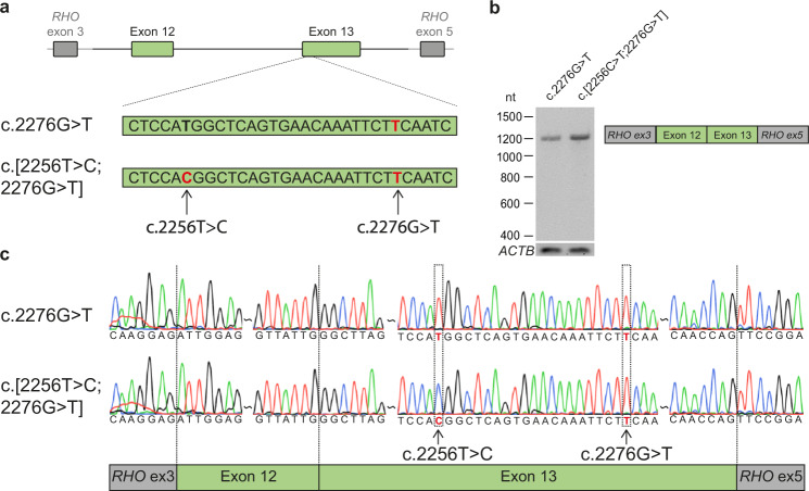
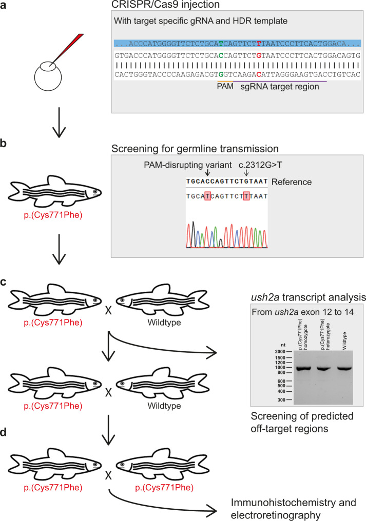
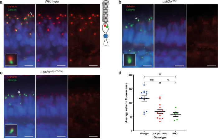
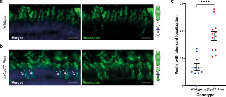
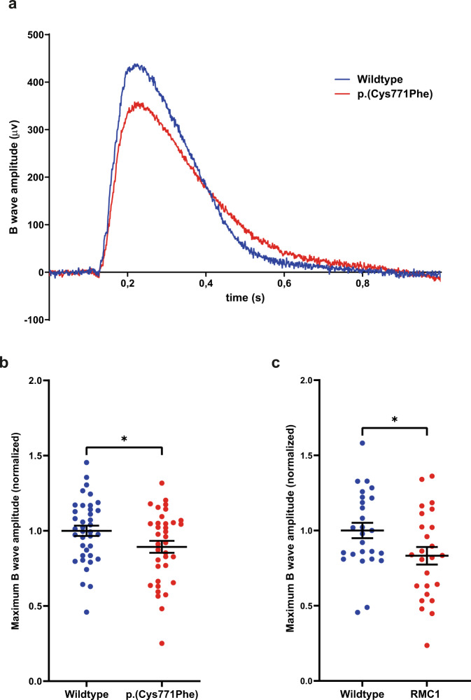
The USH2A variant c.2276 G > T (p.(Cys759Phe)) has been described by many authors as a frequent cause of autosomal recessive retinitis pigmentosa (arRP). However, this is in contrast with the description of two asymptomatic individuals homozygous for this variant. We therefore assessed pathogenicity of the USH2A c.2276 G > T variant using extensive genetic and functional analyses. Whole genome sequencing and optical genome mapping were performed for three arRP cases homozygous for USH2A c.2276 G > T to exclude alternative genetic causes. A minigene splice assay was designed to investigate the effect of c.2276 G > T on pre-mRNA splicing, in presence or absence of the nearby c.2256 T > C variant. Moreover, an ush2ap.(Cys771Phe) zebrafish knock-in model mimicking human p.(Cys759Phe) was generated and characterized using functional and immunohistochemical analyses. Besides the homozygous c.2276 G > T USH2A variant, no alternative genetic causes were identified. Evaluation of the ush2ap.(Cys771Phe) zebrafish model revealed strongly reduced levels of usherin expression at the photoreceptor periciliary membrane, increased levels of rhodopsin localization in the photoreceptor cell body and decreased electroretinogram (ERG) b-wave amplitudes compared to wildtype controls. In conclusion, we confirmed pathogenicity of USH2A c.2276 G > T (p.(Cys759Phe)). Consequently, cases homozygous for c.2276 G > T can now receive a definite genetic diagnosis and can be considered eligible for receiving future QR-421a-mediated exon 13 skipping therapy.


Conflict of interest statement

The authors declare no competing interests.

Figures

Fig. 1
Fig. 1. Minigene splice assays for variants c.2256 C > T and c.2276 G > T.
a A minigene splice assay was performed with a construct spanning from USH2A intron 11 to intron 13 (6,814 nt), containing either c.2276 G > T or both variants (c.[2256 T > C;2276 G > T]). b A single RT-PCR product of 1112 nt was observed after expression of both splice vectors, indicative of the incorporation of USH2A exons 12 and 13 between RHO exons 3 and 5 in both transcripts. c Sanger sequencing confirmed that USH2A exon 12 and exon 13 were correctly incorporated in the mRNA.
Fig. 2
Fig. 2. Generation of the usherinp.(Cys771Phe) zebrafish model.
a Eggs were injected at a one-cell stage with CRISPR/Cas9 mix containing both the target specific single guide RNA (sgRNA) and the 126 nt homology directed repair (HDR) template (partially depicted, in blue) for incorporation of the c.2312 G > T (p.(Cys771Phe)) variant (in red) and protospacer-adjacent motif (PAM, in orange) disrupting variant (c.2304 C > T, in green). The sgRNA target region is depicted in purple. b Once zebrafish were at reproductive age, eggs of the initially injected zebrafish were screened for transmission of the c.2312 G > T and PAM disturbing variant. Three out of ten zebrafish that were screened transmitted the c.2312 G > T variant to their offspring. c Crossbreeding of a c.2312 G > T (p.(Cys771Phe)) zebrafish with a wildtype zebrafish was performed for two generations to reduce unforeseen off-target effects. After the first crossbreeding, genomic DNA was screened for predicted off-target effects of our CRISPR-Cas9 strategy and RNA of homozygous larvae was screened from exon 12 to 14 for deviations on transcript level. d Two p.(Cys771Phe) zebrafish were crossbred with each other to produce homozygous zebrafish. The phenotype of five-day-old larvae was then investigated with immunohistochemistry and electroretinography.
Fig. 3
Fig. 3. Reduced expression level of usherinp.(Cys771Phe) at the photoreceptor periciliary membrane.
a In wildtype zebrafish larval eyes (n = 12 eyes), usherin (red signal) localizes at the photoreceptor periciliary membrane adjacent to the basal body and connecting cilium marker centrin (green signal) as shown by the schematic representation of a photoreceptor on the right. A magnification of one photoreceptor (indicated by an arrow) is depicted in the inlay. b In ush2armc1 knock-out larvae usherin is not detectable (n = 6 eyes). c Localization of usherin at the photoreceptor periciliary membrane was strongly reduced in eyes of ush2ap.(Cys771Phe) larvae (n = 14 eyes) as compared to wildtype. d A Kruskal–Wallis test was performed based on the average of the mean grey value for usherin adjacent to each centrin spot and confirmed a significant decrease of usherin localization adjacent to centrin for the usherinp.(Cys771Phe) and the usherinRMC1 models. The average grey value per retinal section was plotted in a scatter plot (mean ± SEM). Nuclei are stained with DAPI (blue signal). Scale bar: 5 µM. **p: 0.0094, *p: 0.0107, ns not significant.
Fig. 4
Fig. 4. Rhodopsin localization to the photoreceptor cell body in the ush2ap.(Cys771Phe) zebrafish.
a Rhodopsin (green signal) localizes to the rod photoreceptor outer segments in wildtype larval eyes. A visual representation of a photoreceptor is shown on the right. b Aberrant localization of rhodopsin to the photoreceptor cell body was observed in ush2ap.(Cys771Phe) larvae (indicated by white arrows), which was observed in a significantly higher number of photoreceptors of ush2ap.(Cys771Phe) larvae as compared to wildtype larvae (c; unpaired t test). Nuclei are stained with DAPI (blue signal). Scale bar: 10 µM. ****p < 0.0001.
Fig. 5
Fig. 5. Electroretinogram recordings show that ush2ap.(Cys771Phe) mutants are vision impaired.
a Representative electroretinograms of a p.(Cys771Phe) zebrafish and a wildtype sibling at 5 days post-fertilization. b The maximum B wave amplitude was significantly lower in p.(Cys771Phe) zebrafish as compared to wildtype siblings (unpaired t test). The average wildtype amplitude was normalized to 1. Each datapoint corresponds to recordings from an individual larvae (mean ± SEM). *p: 0.0445. c A comparative analysis of ush2armc1 knock-out larvae and age- and strain-matched wildtype larvae was shown to result in a similar and significant decrease in maximum B wave amplitude. Again, the average wildtype amplitude was normalized to 1. Each datapoint corresponds to recordings from an individual larvae (unpaired t test, mean ± SEM). *p: 0.0345.

References

    1. Perea-Romero I, et al. Genetic landscape of 6089 inherited retinal dystrophies affected cases in Spain and their therapeutic and extended epidemiological implications. Sci. Rep. 2021;11:1526. doi: 10.1038/s41598-021-81093-y. - DOI - PMC - PubMed
    1. Stone EM, et al. Clinically focused molecular investigation of 1000 consecutive families with inherited retinal disease. Ophthalmology. 2017;124:1314–1331. doi: 10.1016/j.ophtha.2017.04.008. - DOI - PMC - PubMed
    1. Jouret G, et al. Genetics of Usher syndrome: new insights from a meta-analysis. Otol. Neurotol. 2019;40:121–129. doi: 10.1097/MAO.0000000000002054. - DOI - PubMed
    1. Toualbi L, Toms M, Moosajee M. USH2A-retinopathy: from genetics to therapeutics. Exp. Eye Res. 2020;201:108330. doi: 10.1016/j.exer.2020.108330. - DOI - PMC - PubMed
    1. Rivolta C, Sweklo EA, Berson EL, Dryja TP. Missense mutation in the USH2A gene: association with recessive retinitis pigmentosa without hearing loss. Am. J. Hum. Genet. 2000;66:1975–1978. doi: 10.1086/302926. - DOI - PMC - PubMed

LinkOut - more resources