. 2022 Jun 7;12(1):9362.
doi: 10.1038/s41598-022-13257-3.

First ovum-in-ovo pathological titanosaurid egg throws light on the reproductive biology of sauropod dinosaurs

Affiliations

First ovum-in-ovo pathological titanosaurid egg throws light on the reproductive biology of sauropod dinosaurs

Harsha Dhiman et al. Sci Rep. .

Abstract

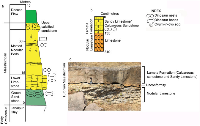
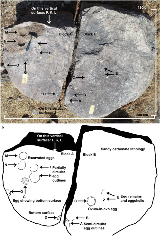
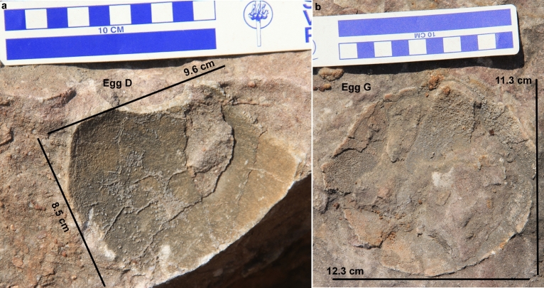
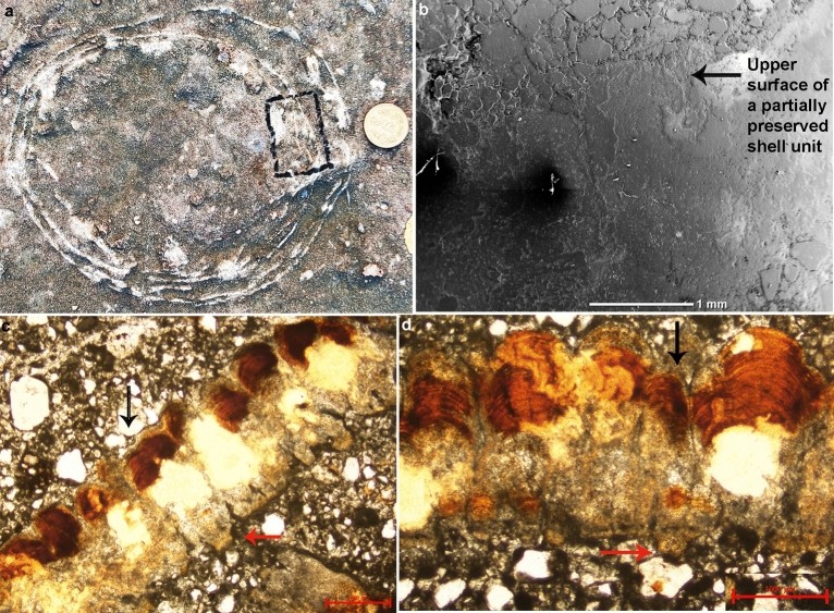
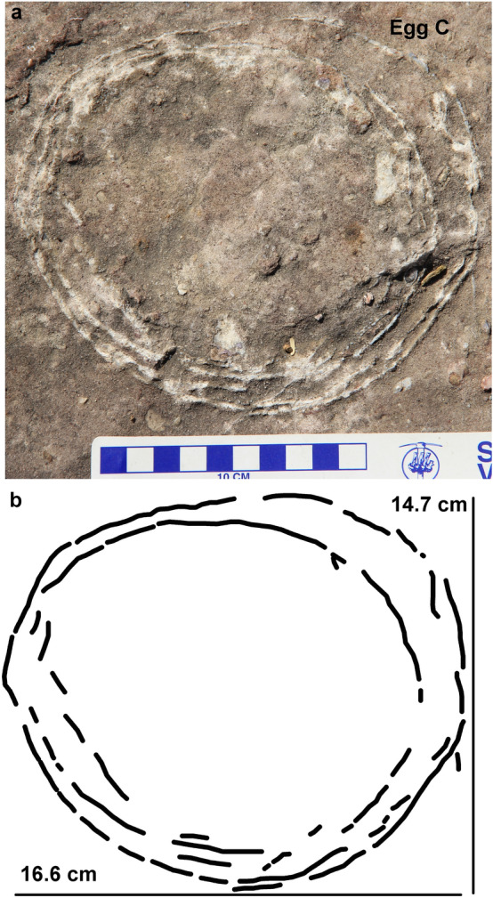
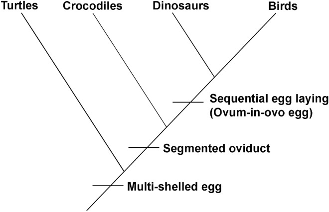
Pathologic eggs have been documented in the amniote eggs of birds, turtles, and dinosaurs. These eggs occur either in the form of one egg within another egg, a condition known as ovum-in-ovo or multi-shelled eggs showing additional pathological eggshell layer/s besides the primary shell layer. Though multi-shelled eggs and eggshells were previously recorded only in reptiles and ovum-in-ovo eggs in birds, now it has been shown that multi-shelled egg pathology occurs in birds as well. However, no ovum-in-ovo egg has been reported in dinosaurs or for that matter in other reptiles. Here we describe an ovum-in-ovo pathological egg from a titanosaurid dinosaur nest from the Upper Cretaceous Lameta Formation of western Central India which makes it the first report of this pathology in dinosaurs. Birds possess a specialized uterus while other amniotes have a generalized uterus. However, alligators and crocodiles retain a specialized uterus like birds along with a reptilian mode of egg-laying. The discovery of ovum-in-ovo egg from a titanosaurid dinosaur nest suggests that their oviduct morphology was similar to that of birds opening up the possibility for sequential laying of eggs in this group of sauropod dinosaurs. This new find underscores that the ovum-in-ovo pathology is not unique to birds and sauropods share a reproductive behavior very similar to that of other archosaurs.


Conflict of interest statement

The authors declare no competing interests.

Figures

Figure 1
Figure 1
Pathologic cledoic eggs showing unusual morphology not similar to the  normal eggs. (a) Ovum-in-ovo pathologic egg characterized by an egg within an egg or two yolks. (b) Multi-shelled egg distinctive of two or more than two eggshell layers surrounding an individual egg (modified after Carpenter, software used Adobe Illustrator CS6).
Figure 2
Figure 2
Location map of the study area showing pathologic dinosaur nest P7 from the village Padlya, Dhar District, Madhya Pradesh, India (software used Adobe Illustrator CS6).
Figure 3
Figure 3
Stratigraphic subdivisions of the Lameta Formation; (a) Jabalpur (modified after Tandon et al.); (b) Padlya; (c) Field photograph showing the  sandy  limestone/calcareous  sandstone of the Lameta Formation overlying the Nodular Limestone of the Bagh Group within Dinosaur Fossil National Park (DFNP), near Padlya, Dhar district, M.P. The Nodular Limestone (Bagh Beds) shows lensoid character while the sandy limestone/calcareous sandstone has massive appearance (a,b drawn using software Adobe Photoshop CC).
Figure 4
Figure 4
Field photograph and schematic diagram of the outcrop showing nest P7 and its eggs and eggshell fragments belonging to a titanosaur sauropod randomly spaced with respect to each other in a circular nest arrangement. The captions A to O indicate eggs and eggshell locations (Fig. 4b drawn using software Adobe Photoshop CC).
Figure 5
Figure 5
Field photograph of eggs A and B from nest number P7. The eggs exist as semi-circular outlines and the gaps (see arrows) possibly represent hatching windows.
Figure 6
Figure 6
Field photographs of eggs from nest number P7. (a) Circular rock with few encrusted eggshells. (b) Elliptical  egg with shell fragment pile. (c) Circular to semi-circular eggs.
Figure 7
Figure 7
Field photographs of eggs from nest number P7. (a) Partially preserved bottom surface of an egg. (b) Circular bottom surface of an egg with encrusted eggshells.
Figure 8
Figure 8
Field photographs of eggs from nest number P7 showing randomly distributed eggshell fragments.
Figure 9
Figure 9
Field photograph and microscopic images of the eggshell from egg number C showing oospecies Fusioolithus baghensis characterized by fan-shaped shell units with arching growth lines and basal end cap units. (a) The ovum-in-ovo egg with the boxed area showing the region from where the specimen was extracted. (b) Radial Scanning Electron Microscope (SEM) photomicrograph of the eggshell showing partially preserved fan-shaped shell unit. (c) Photomicrograph of the radial thin section of the eggshell under polarized light microscopy showing partially preserved shell units (see black arrow) and characteristic swollen basal cap units (see red arrow). (d) Photomicrograph of the radial thin section of the eggshell under polarized light microscopy exhibiting fused shell units and growth lines (see black arrow) and swollen basal cap (see red arrow).
Figure 10
Figure 10
Field photograph and microscopic images of the eggshells from egg number M showing oospecies Fusioolithus baghensis characterized by fan-shaped shell units with arching growth lines and basal end cap units. (a) Egg numbers M and N present as sub-circular outlines. The eggs were excavated and eggshells were studied for parataxonomic classification. (b) X-Ray Microscopy photomicrograph of the eggshell showing fan-shaped shell units progressing upwards into bumpy nodes. Arching growth lines restricted to shell units can also be seen. (c) Radial SEM photograph showing curvy growth lines in the middle of the shell unit. (d) Radial thin section showing fan-shaped shell units merging with each other in the right segment where growth lines exist as sub-horizontal lines and partially preserved basal end units.
Figure 11
Figure 11
Field photograph and schematic diagram of the pathologic ovum-in-ovo egg (egg number C) documented from the Lameta Formation of village Padlya, Dhar District, M.P., India. Two slightly partially broken circular eggshell outlines can be seen with broken eggshell fragments within. A crescent-shaped gap is characteristically present in the upper right part of the egg (b drawn using software Adobe Photoshop CC).
Figure 12
Figure 12
Inferred cladogram showing divergence of dinosaurs from crocodiles on the basis of sequential egg laying (figure drawn using software Adobe Photoshop CC).

Similar articles

Cited by

References

    1. Sander PM. Reproduction in early amniotes. Science. 2012;337(6096):806–808. doi: 10.1126/science.1224301. - DOI - PubMed
    1. Norell MA, et al. A theropod dinosaur embryo and the affinities of the Flaming Cliffs dinosaur eggs. Science. 1994;266(5186):779–782. doi: 10.1126/science.266.5186.779. - DOI - PubMed
    1. Carpenter K. Eggs, Nests, and Baby Dinosaurs: A Look at DINOSAUR Reproduction. Indiana University Press; 1999.
    1. Horner JR. Dinosaur reproduction and parenting. Annu. Rev. Earth Planet. Sci. 2000;28(1):19–45. doi: 10.1146/annurev.earth.28.1.19. - DOI
    1. Chiappe, L. M., Jackson, F., Coria, R. A. & Dingus, L. Nesting titanosaurs from Auca Mahuevo and adjacent sites. In The Sauropods, 285–302 (University of California Press, 2005).

Publication types