. 2022 Jun 7;22(1):26.
doi: 10.1186/s40644-022-00464-4.

Does the addition of whole-body MRI to routine imaging influence real-world treatment decisions in metastatic breast cancer?

Affiliations

Does the addition of whole-body MRI to routine imaging influence real-world treatment decisions in metastatic breast cancer?

Basrull N Bhaludin et al. Cancer Imaging. .

Abstract

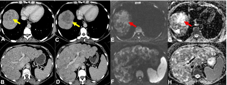
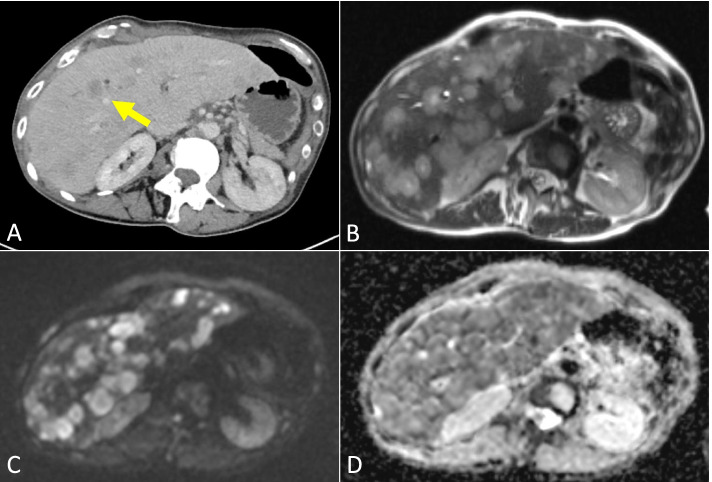
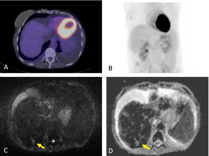
Background: The assessment of metastatic breast cancer (MBC) can be limited with routine imaging such as computed tomography (CT) especially in bone-only or bone-predominant disease. This analysis investigates the effects of the use of WBMRI in addition to the use of routine CT, bone scintigraphy (BS) and fluorine-18-fluorodeoxyglucose positron emission tomography with computed tomography (FDG-PET/CT) on influencing systemic anti-cancer treatment (SACT) decisions in patients with known MBC.

Methods: MBC patients undergoing SACT who had WBMRI undertaken within 8 weeks of either a routine CT, BS or FDG-PET/CT were reviewed retrospectively. The clinical indications for undertaking the WBMRI examinations were recorded. Data on the extent and distribution of the disease were collected and discordance/concordance of disease status across the imaging modalities were compared. SACT decisions at each time point were also evaluated.

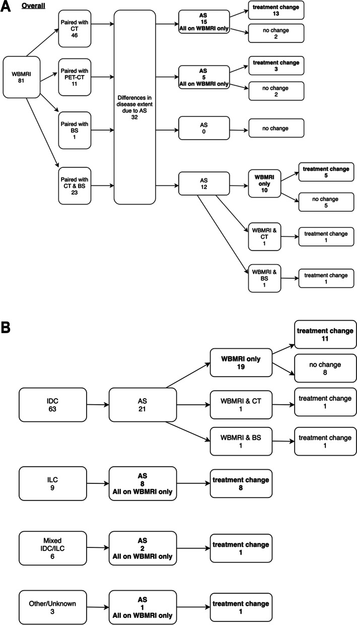
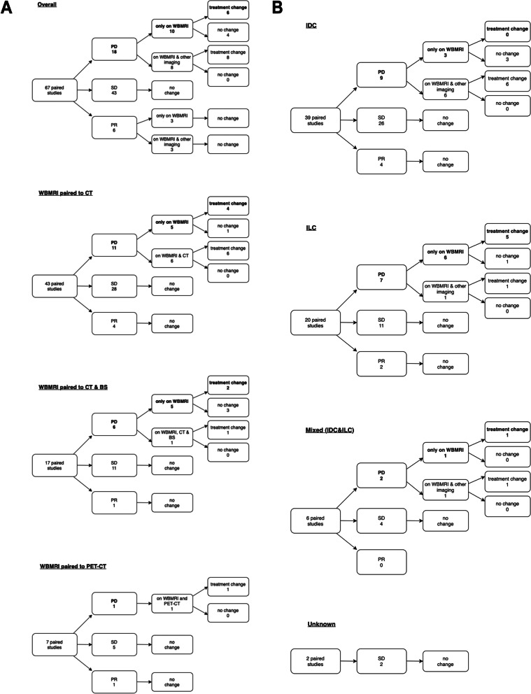
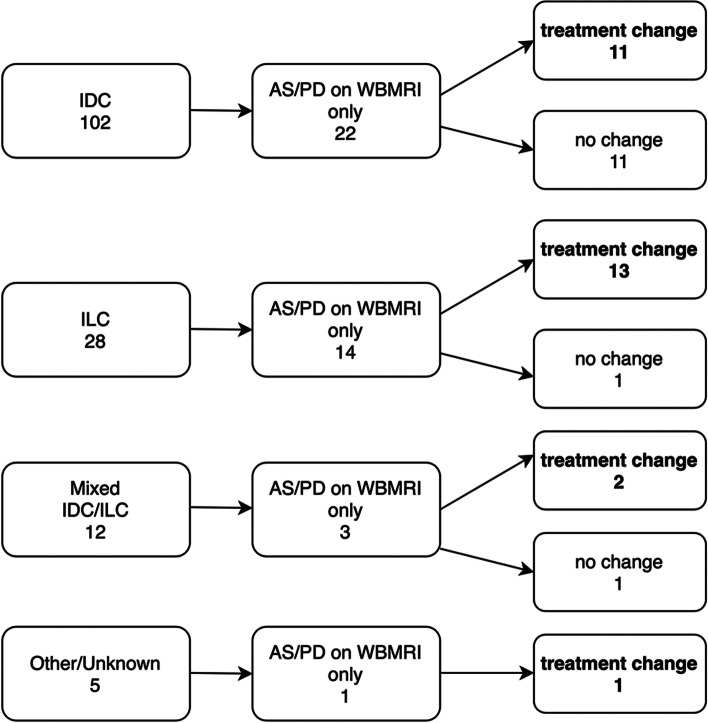
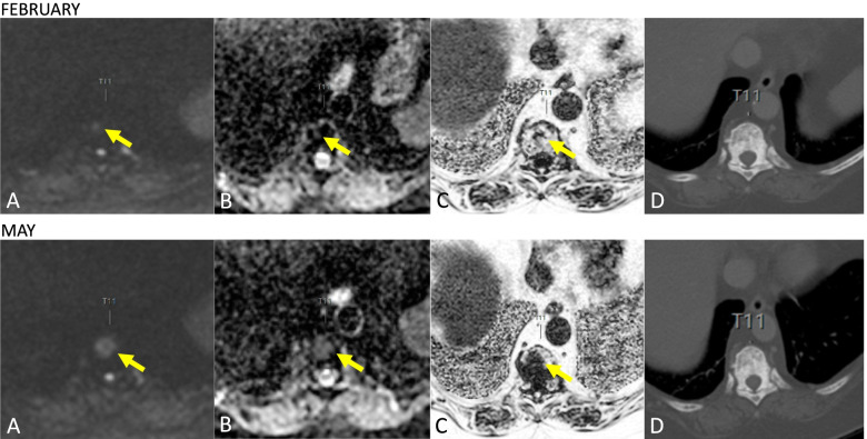
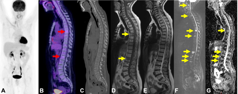
Results: There were 105 MBC patients with 148 WBMRI studies paired with CT, BS or FDG-PET/CT. 50 pairs (33.8%) showed differences in the extent of disease, with 44 pairs due to additional sites (AS) reported on WBMRI alone. 81 patients (Group 1) had one WBMRI paired with routine imaging due to a variety of indications, with clinical symptoms (such as bone pain) being the most common (24.7%). 24 patients (Group 2) had more than one WBMRI study paired with routine imaging comprising 67 pairs. 13/67 pairs (19.4%) showed discordance in assessments. 10/13 pairs had progressive disease (PD) reported on WBMRI alone. SACT change due to AS reported on WBMRI alone occurred in 21/23 pairs (91.3%) in Group 1. SACT change due to PD reported on WBMRI alone in Group 2 occurred in 6/14 pairs (42.9%). SACT change due to AS/PD in both groups occurred in 11/102 pairs (10.8%) with known invasive ductal carcinoma (IDC) and 13/28 pairs (46.4%) with invasive lobular carcinoma (ILC).

Conclusions: The use of WBMRI in MBC led to earlier recognition of PD and SACT change compared with the other imaging modalities. A higher proportion of discordant response assessments and SACT changes were observed in ILC compared with IDC in our patient group, although larger-scale studies are required to investigate this further.

Keywords: Cancer treatment; Diffusion-weighted imaging; Metastatic breast cancer; Response assessment; Whole-body magnetic resonance imaging.


Conflict of interest statement

The authors declare that they have no competing interests.

Figures

Fig. 1
Fig. 1
WBMRI better characterising diffuse liver disease. 43-year-old female with patient with ER- PR- positive HER-2 negative metastatic breast cancer to the nodes, lung and liver was being monitored with CT during chemotherapy treatment. Follow up CT following 3 cycles demonstrates enlarging and more confluent liver lesion at the liver dome (C – yellow arrow) compared to prior CT (A—yellow arrow) and a greater number of lesions elsewhere within the liver suggesting worsening disease (D) when compared to prior CT (B) however tumour markers were improving on treatment. The increasingly irregular liver contour suggests pseudocirrhosis (D). WBMRI was performed to further evaluate the CT findings. The high b value (b900) DWI images (E) and corresponding ADC map (F) show diffuse disease with some lesions demonstrating high ADC values representing low cellularity/treated disease and others demonstrating low ADC values representing high cellularity/active disease in keeping with a mixture of treated and active disease, indicating a favourable response to treatment in some areas
Fig. 2
Fig. 2
a Flowchart showing the different combinations of paired examinations, the distribution of the differences in disease extent and treatment changes in Group 1. b Flowchart showing the distribution of additional sites of disease and treatment changes according to histological subtypes in Group 1
Fig. 3
Fig. 3
a Flowcharts showing the differences in response assessments between the paired imaging modalities and treatment changes in Group 2. b Flowcharts showing the differences in response assessments between the paired imaging modalities and treatment changes in Group 2 according to histological subtypes
Fig. 4
Fig. 4
Pooled data on the number of AS/PD reported on WBMRI only and treatment changes according to histological subtypes
Fig. 5
Fig. 5
Disease underestimated by CT. 55-year-old female with ER- PR- positive HER-2 negative infiltrating ductal carcinoma with axillary nodal and liver metastases. CT demonstrates bi-lobar liver metastases but significantly underestimates the burden of disease when compared to the WBMRI performed 2 days later. CT (A) demonstrates liver metastases measuring up to 18 mm within segment VIII (yellow arrow) however axial T2 (B), high b-value (b900) DWI sequences (C) and corresponding ADC map (D) from the WBMRI study demonstrates many more lesions not appreciated on CT
Fig. 6
Fig. 6
Additional sites of disease on WBMRI compared to FDG/PET-CT. 56-year-old female with ER- PR- positive HER-2 negative infiltrating ductal carcinoma with bone and liver metastases. Worsening liver function tests with decreasing haemoglobin and platelet count were noted whilst on Exemestane. Fused PET-CT images (A) and FDG/PET-CT MIP (B) showed no significant pathological activity in the liver. There is pseudocirrhosis, ascites and pleural effusions. High b-value (b900) image (C) and corresponding ADC map (D) of a WBMRI study undertaken four weeks later demonstrated multiple liver metastases measuring up to 20 mm in the posterior right hepatic lobe (yellow arrow) not appreciated on the FDG/PET-CT study
Fig. 7
Fig. 7
Progressive disease on WBMRI not identified on CT. 80-year-old female with ER- PR- positive HER-2 negative infiltrating ductal carcinoma with bone metastases on first presentation. Tumour marker rise was noted whilst on Letrozole and Denusomab over a three-month period. WBMRI demonstrated clear progression at several sites but no changes were appreciated on CT performed at the same time points. In this figure high b-value (b900) image (A), corresponding ADC map (B) and derived fat fraction images from the Dixon sequences (C) demonstrate an increase in the size of the lesion at T11 with no appreciable change in the mixed lytic and sclerotic disease burden on CT (D)
Fig. 8
Fig. 8
FDG PET-CT and WB MRI in lobular breast cancer. 62-year-old female with a history of treated lobular breast cancer with no history of metastatic disease presented with back pain. FDG PET-CT MIP (A) showed no significant or high grade pathological activity. The fused PET-CT images (B) showed minimal activity in a few bone lesions (red arrows) although the lesions were barely appreciated on the low-dose CT component (C). These were indeterminate but suspicious in this context. WBMRI was performed for further evaluation which showed a greater number of bone lesions (yellow arrows) better seen on Sagittal T1-weighted images (D) compared with the Sagittal T2-weighted images (E). The bone lesions demonstrate restricted diffusion with high signal on b900 DWI images (F) and low signal on ADC map (G) in keeping with bone metastases

References

    1. The Royal College of Radiologists. Guidance on screening and symptomatic breast imaging: Fourth edition. Clinical Radiology The Royal College of Radiologists [Internet]. The Royal College of Radiologists; 2019 [cited 2021 May 9]; Available from: www.rcr.ac.uk
    1. Gradishar WJ, Moran MS, Abraham J, Aft R, Agnese D, Allison KH, et al. NCCN Guidelines® insights: breast cancer, Version 4.2021. J Natl Comprehens Cancer Netw. 2021;19(484):93. - PubMed
    1. Gennari A, André F, Barrios CH, Cortés J, de Azambuja E, DeMichele A, et al. ESMO Clinical Practice Guideline for the diagnosis, staging and treatment of patients with metastatic breast cancer☆. Ann Oncol. 2021;32:1475–1495. doi: 10.1016/j.annonc.2021.09.019. - DOI - PubMed
    1. Eisenhauer EA, Therasse P, Bogaerts J, Schwartz LH, Sargent D, Ford R, et al. New response evaluation criteria in solid tumours: Revised RECIST guideline (version 1.1) Eur J Cancer. 2009;45:228–47. doi: 10.1016/j.ejca.2008.10.026. - DOI - PubMed
    1. Coleman RE. Clinical Features of Metastatic Bone Disease and Risk of Skeletal Morbidity. Clin Cancer Res. 2006;12(6243s LP):6249s. - PubMed

MeSH terms

Substances