

Left Main Coronary Artery Fusiform Aneurysm
- PMID: 35673313
- PMCID: PMC9165914
- DOI: 10.7759/cureus.24790
Left Main Coronary Artery Fusiform Aneurysm
Abstract
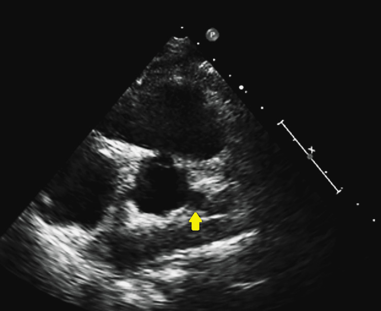
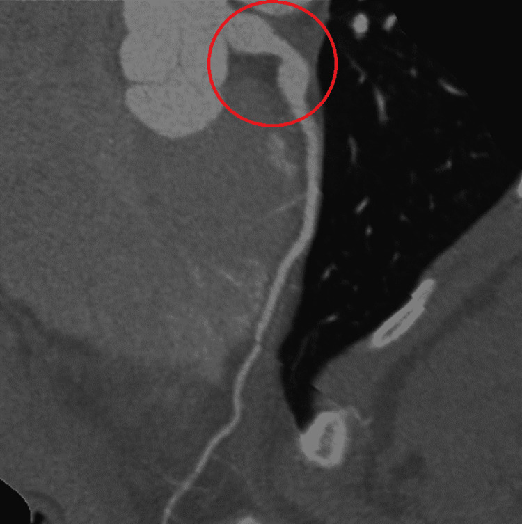
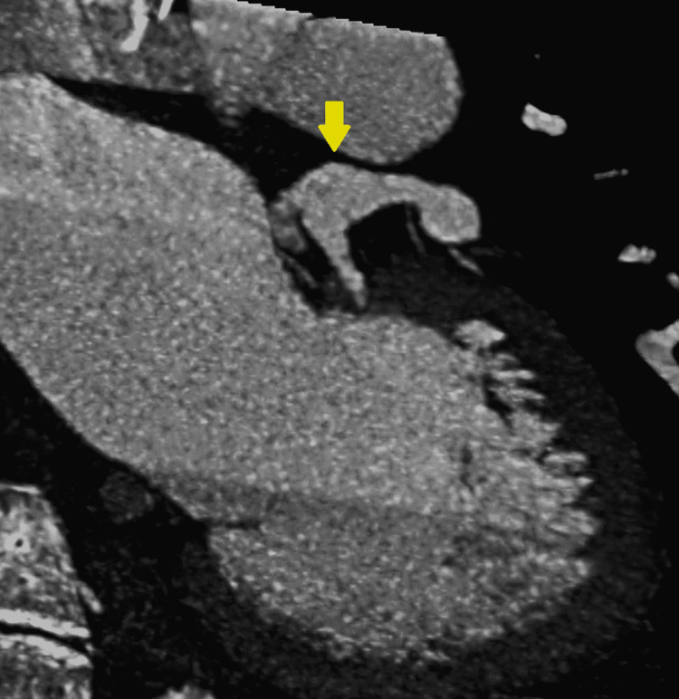
Aneurysm of the coronary artery is an uncommon condition that is usually found incidentally. The left coronary aneurysm is the least common. We report the case of a young patient with a history of vasculitis who was found to have a left fusiform coronary aneurysm. This is a 20-year-old female who has a history of polyarteritis nodosa and who came due to shortness of breath associated with chest discomfort. The physical exam was only relevant for multiple joint pains and tenderness. An echocardiogram showed a possible coronary aneurysm that was confirmed on the angio-tomography. The patient was discharged without complications. The left main coronary artery aneurysm is a rare condition and the least common of the coronary aneurysms. There is no established guideline for screening and therapy of these aneurysms, but invasive methods are not a preferred method for follow-up on this condition.
Keywords: coronary aneurysm; fusiform coronary aneurysm; left main coronary artery aneurysm; polyarteritis nodosom; vasculitis.
Copyright © 2022, Rodriguez Guerra et al.
Conflict of interest statement
The authors have declared that no competing interests exist.
Figures
Similar articles
-
Giant coronary artery aneurysms involving more than one coronary artery: case report.J Cardiothorac Surg. 2021 Jun 19;16(1):177. doi: 10.1186/s13019-021-01560-5. J Cardiothorac Surg. 2021. PMID: 34147120 Free PMC article.
-
Left main coronary artery stenosis associated with extremely long fusiform aneurysm.Int J Cardiol. 2007 Sep 14;121(1):97-9. doi: 10.1016/j.ijcard.2006.08.038. Epub 2006 Nov 13. Int J Cardiol. 2007. PMID: 17107722
-
An Incidentally Discovered Large Left Main Coronary Artery Aneurysm.Cureus. 2020 Jul 13;12(7):e9172. doi: 10.7759/cureus.9172. Cureus. 2020. PMID: 32802608 Free PMC article.
-
Insidious coronary artery disease in a young patient with polyarteritis nodosa: a case report and literature review.BMC Cardiovasc Disord. 2021 Feb 27;21(1):115. doi: 10.1186/s12872-021-01923-9. BMC Cardiovasc Disord. 2021. PMID: 33639843 Free PMC article. Review.
-
Endovascular Management of Multiple Dysplastic Aneurysms in a Young Man with an Unknown Underlying Cause: A Case Report and Review of the Literature.World Neurosurg. 2018 Mar;111:349-357. doi: 10.1016/j.wneu.2017.12.129. Epub 2017 Dec 28. World Neurosurg. 2018. PMID: 29289804 Review.
References
-
- Aneurysm of the coronary arteries. Packard M, Wechsler HF. Arch Intern Med. 1929;43:1–14.
-
- Aneurysmal coronary artery disease. Swaye PS, Fisher LD, Litwin P, et al. Circulation. 1983;67:134–138. - PubMed
-
- Coronary artery aneurysms: a review of the natural history, pathophysiology, and management. Cohen P, O'Gara PT. Cardiol Rev. 2008;16:301–304. - PubMed
-
- New-onset coronary aneurism and late-acquired incomplete scaffold apposition after full polymer jacket of a chronic total occlusion with bioresorbable scaffolds. Gargiulo G, Mangiameli A, Granata F, et al. JACC Cardiovasc Interv. 2015;8:41–43. - PubMed
Publication types
LinkOut - more resources
Full Text Sources