Case Reports
. 2022 Jun;81(6):151-154.

A Rare Presentation of Central Nervous System Tuberculomas in an Immunocompetent Patient

Affiliations
Case Reports

A Rare Presentation of Central Nervous System Tuberculomas in an Immunocompetent Patient

Bryce K Chang et al. Hawaii J Health Soc Welf. 2022 Jun.

Abstract

The uncommon presentation of simultaneous brain and lung lesions in an immunocompetent adult patient with frequent travel to a mycobacterium tuberculosis (MTB) endemic area requires high clinical suspicion for central nervous system (CNS) MTB, as this disease often results in severe neurologic morbidity and mortality. Non-specific and subacute symptoms make the diagnosis of CNS MTB clinically challenging, and a workup with imaging and microbiological studies such as acid-fast bacilli staining, nucleic acid amplification testing, and tissue culture must not delay prompt treatment with anti-tuberculosis therapy. This case illustrates the complex challenges of medical diagnosis and multi-disciplinary decision-making involved in the workup of CNS MTB.

Keywords: central nervous system; mycobacterium tuberculosis; simultaneous brain and lung lesions.


Figures

Figure 1.
Figure 1.
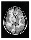
Multiple areas of increased T2 signal on the T2 FLAIR sequences throughout both hemispheres with right to left midline shift of approximately 3 mm.
Figure 2.
Figure 2.
MRI showing innumerable ring enhancing masses throughout the brain parenchyma with extensive vasogenic edema with largest mass measuring 2.0 × 1.4 × 1.2 cm in the right temporoparietal region.
Figure 3.
Figure 3.
Non-contrast chest CT significant for bi-apical consolidations with air bronchograms suspicious for tuberculosis.

References

    1. Hawaii Tuberculosis Control Program State of Hawaii. 2021. Updated July 1, 2020. https://health.hawaii.gov/tb/data-statistics/
    1. Thwaites G, Fisher M, Hemingway C, Scott G, Solomon T, Innes J. British Infection Society guidelines for the diagnosis and treatment of tuberculosis of the central nervous system in adults and children. J Infect. 2009;59((3)):167–187. doi: 10.1016/j.jinf.2009.06.011. - DOI - PubMed
    1. Schaller MA, Wicke F, Foerch C, Weidauer S. Central Nervous System Tuberculosis : etiology, clinical manifestations and neuroradiological features. Clin Neuroradiol. 2019;29((1)):3. doi: 10.1007/s00062-018-0726-9. - DOI - PubMed
    1. Bahr NC, Meintjes G, Boulware DR. Inadequate diagnostics: the case to move beyond the bacilli for detection of meningitis due to Mycobacterium tuberculosis. J Med Microbiol. 2019;68((5)):755. doi: 10.1099/jmm.0.000975. - DOI - PMC - PubMed
    1. Cheng VCC, Yam WC, Hung IFN, et al. Clinical evaluation of the polymerase chain reaction for the rapid diagnosis of tuberculosis. J Clin Pathol. 2004;57((3)):281. doi: 10.1136/jcp.2003.012658. - DOI - PMC - PubMed

Publication types