. 2022 May 13;12(9):4110-4126.
doi: 10.7150/thno.71392. eCollection 2022.

Wnt4 is crucial for cardiac repair by regulating mesenchymal-endothelial transition via the phospho-JNK/JNK

Affiliations

Wnt4 is crucial for cardiac repair by regulating mesenchymal-endothelial transition via the phospho-JNK/JNK

Wenyan Dong et al. Theranostics. .

Abstract

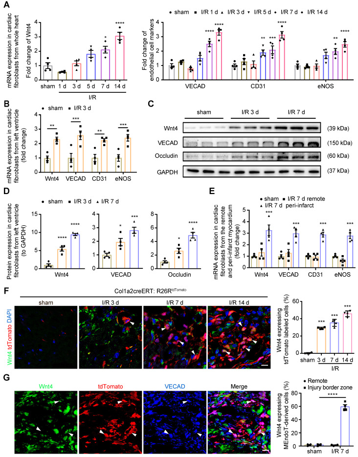
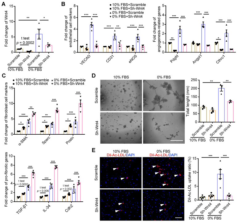
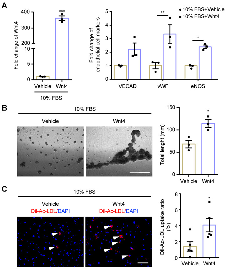
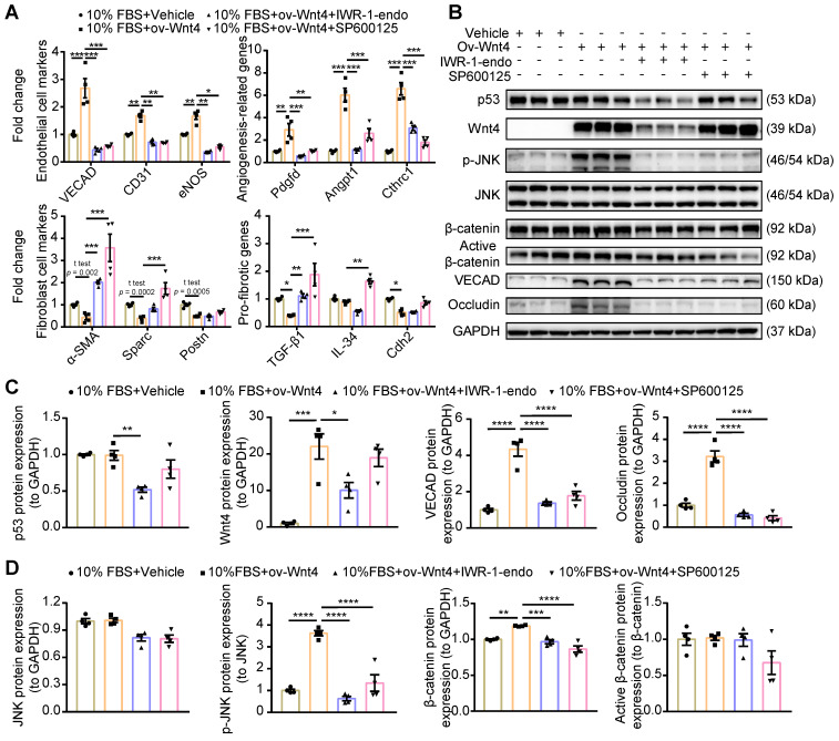
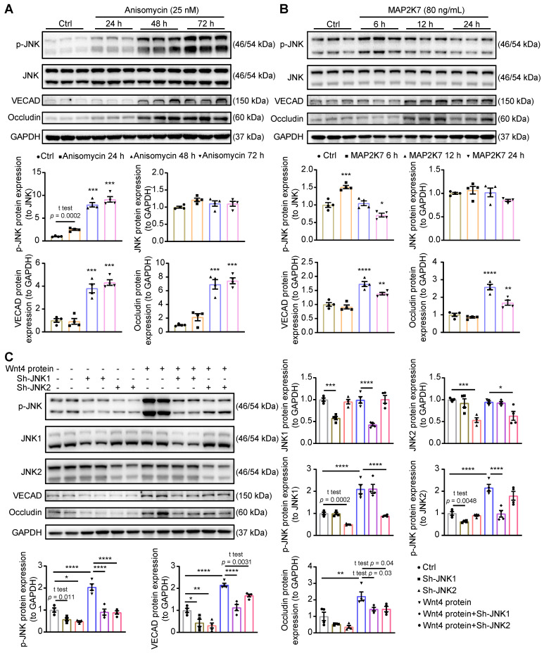
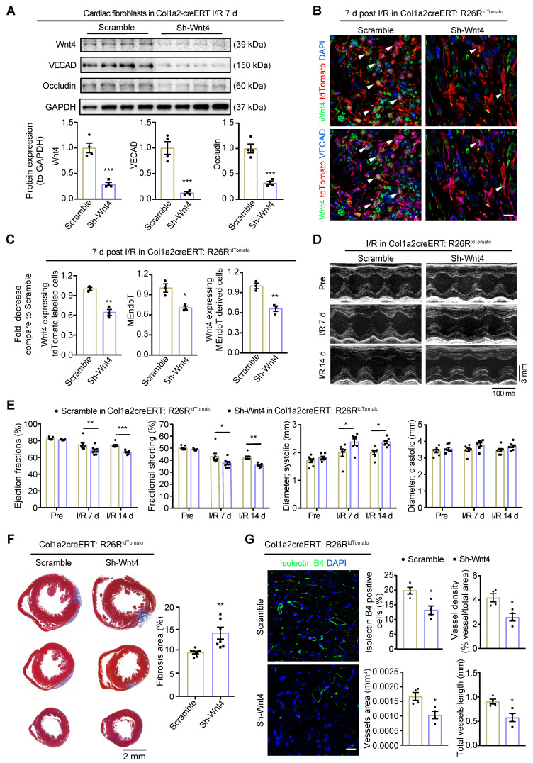
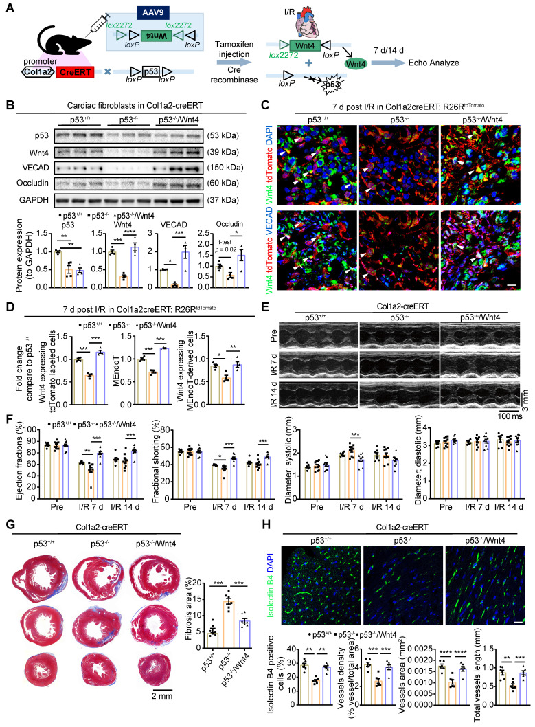
Rational: Wnt4 plays a critical role in development and is reactivated during fibrotic injury; however, the role of Wnt4 in cardiac repair remains unclear. In this study, our aim was to clarify the pathophysiological role and mechanisms of Wnt4 following acute cardiac ischemic reperfusion injury. Methods and results: We investigated the spatio-temporal expression of Wnt4 following acute cardiac ischemic reperfusion injury and found that Wnt4 was upregulated as an early injury response gene in cardiac fibroblasts near the injury border zone and associated with mesenchymal-endothelial transition (MEndoT), a beneficial process for revascularizing the damaged myocardium in cardiac repair. Using ChIP assay and in vitro and in vivo loss- and gain-of-function, we demonstrated that Wnt4 served as a crucial downstream target gene of p53 during MEndoT. Wnt4 knockdown in cardiac fibroblasts led to decreased MEndoT and worsened cardiac function. Conversely, Wnt4 overexpression in cardiac fibroblasts induced MEndoT in these cells via the phospho-JNK/JNK signaling pathway; however, both the p53 and Wnt4 protein levels were dependent on the β-catenin signaling pathway. JNK activation plays a critical role in the induction of MEndoT and is crucial for Wnt4 regulated MEndoT. Moreover, Wnt4 overexpression specifically in cardiac fibroblasts rescued the cardiac function worsening due to genetic p53 deletion by decreasing fibrosis and increasing MEndoT and vascular density. Conclusion: Our study revealed that Wnt4 plays a pivotal role in cardiac repair with involvement of phospho-JNK mediated MEndoT and is a crucial gene for cardiac fibroblast-targeted therapy in heart disease.

Keywords: Cardiac ischemic reperfusion injury; Wnt4; cardiac fibroblast; mesenchymal-endothelial transition (MEndoT); p53.


Conflict of interest statement

Competing Interests: The authors have declared that no competing interest exists.

Figures

Figure 1
Figure 1
Wnt4 upregulated in cardiac fibroblasts after acute ischemic reperfusion (I/R) injury. (A) qPCR analysis of Wnt4 and endothelial cell marker genes in cardiac fibroblasts isolated within 24 h from whole heart tissue, n = 4. (B) qPCR analysis of Wnt4 and endothelial cell marker genes in cardiac fibroblasts isolated within 24 h from the left ventricular heart tissue early at day 3 post I/R injury, n = 4. (C-D) Western blot for Wnt4 and endothelial cell marker genes in cardiac fibroblasts isolated within 24 h from the left ventricular heart tissue post I/R injury and the densitometric quantification, n = 4. (E) qPCR analysis of Wnt4 and endothelial cell marker genes in cardiac fibroblasts respectively isolated within 24 h from the remote and peri-infarct myocardium, n = 4. (F) Immunofluorescence staining of Wnt4 in injury border zone of heart section from Col1a2-CreERT: R26RtdTomato mice post sham or I/R injury and the quantification, n = 4. (G) Triple immunofluorescence staining of tdTomato, Wnt4 and VECAD in injury border zone and the quantification, n = 4. (All graphs show mean ± S.E.M; *p < 0.05, **p < 0.01, ***p < 0.001, ****p < 0.0001, using one-way anova and two-way anova compared with sham. Colocalization of fluorophores is indicated by arrowhead. Scale bar: 10 µm).
Figure 2
Figure 2
p53 regulate MEndoT by directly targeting Wnt4 promotor regions. (A) qPCR analysis of Wnt4 and endothelial cell marker genes in cardiac fibroblasts after serum starvation and treated with RITA (p53 activator, 50 µM) and pifithrin-α (p53 inhibitor, 50 µM), n = 4. (B) The chromatin immunoprecipitation (ChIP) sequencing for p53 on serum-starved cardiac fibroblasts showed that Wnt4 promoter region was a direct-binding site of p53. (C) ChIP-qPCR assay of Wnt4 after p53 ChIP, n = 3. (D) The enhanced MEndoT by RITA was inhibited after Wnt4 knockdown, n = 4. (All graphs show mean ± S.E.M; *p < 0.05, **p < 0.01, ***p < 0.001, using unpaired t-test, one-way anova and two-way anova). Note: FBS (fetal bovine serum), RITA (reactivation of p53 and induction of tumour apoptosis).
Figure 3
Figure 3
MEndoT downregulated by Wnt4 knockdown. (A-C) qPCR analysis of gene expression in cardiac fibroblasts after Wnt4 shRNA lentivirus transduction, n = 4. (A) Wnt4. (B) Endothelial cell marker genes and angiogenesis-related genes. (C) Fibroblast cell marker genes and pro-fibrotic genes. (D) Capillary tube formation on Matrigel and the quantification, n = 3, Scale bar: 750 µm. (E) Ac-LDL intake and the quantification, n = 5, Scale bar: 200 µm. (All graphs show mean ± S.E.M; *p < 0.05, **p < 0.01, ***p < 0.001, using unpaired t-test, one-way anova and two-way anova, fluorophores is indicated by arrowhead.) Note: Angpt1 (angiopoietin 1), α-SMA (actin alpha 2, smooth muscle, aorta), CD31 (Pecam1, platelet and endothelial cell adhesion molecule 1), Cdh2 (cadherin 2), Cthrc1 (collagen triple helix repeat containing 1), eNOS (nitric oxide synthase 3), IL-34 (interleukin 34), Pdgfd (platelet derived growth factor D), Postn (periostin), Sparc (secreted acidic cysteine rich glycoprotein), TGF-β1 (transforming growth factor beta 1), VECAD (cadherin 5).
Figure 4
Figure 4
MEndoT can be induced by Wnt4 overexpression. The cardiac fibroblasts were transducted with Wnt4 lentivirus and cultured in 10% FBS cell culture conditions. (A) qPCR analysis of Wnt4 and endothelial cell markers, n = 3. (B) Capillary tube formation on Matrigel and the quantification, n = 3, Scale bar: 750 µm. (C) Ac-LDL intake and the quantification, n = 5, Scale bar: 200 µm. (All graphs show mean ± S.E.M; *p < 0.05, **p < 0.01, ***p < 0.001, using unpaired t-test and two-way anova, fluorophores is indicated by arrowhead.)
Figure 5
Figure 5
Wnt4 induce MEndoT via the p-JNK/JNK signaling pathway. (A) qPCR analysis of gene expression after Wnt4 overexpression in cardiac fibroblasts to induce MEndoT and added β-catenin inhibitors IWR-1-endo (50 µM) and p-JNK/JNK pathway inhibitor SP600125 (50 µM). (B-D) Western blot for cardiac fibroblasts and the quantification. (All graphs show mean ± S.E.M; *p < 0.05, **p < 0.01, ***p < 0.001, ****p < 0.0001, n = 4, using unpaired t-test, one-way anova and two-way anova.)
Figure 6
Figure 6
Activation of JNK is crucial for Wnt4 protein induced MEndoT. (A) Western blot analysis of the expression of p-JNK, JNK, VECAD and Occludin in cardiac fibroblasts after adding with anisomycin (JNK activator, 25 nM) for 24 h, 48 h and 72 h, and the quantification. n = 4. (B) Western blot analysis of the expression of p-JNK, JNK, VECAD and Occludin in cardiac fibroblasts after adding with recombinant MAP2K7 (JNK kinase, 80 ng/mL) for 6 h, 12 h and 24 h, and the quantification. n = 4. (C) Western blot analysis of the expression of p-JNK, JNK1, JNK2, VECAD and Occludin in cardiac fibroblasts, and the quantification. JNK1 or JNK2 were knock down in cardiac fibroblasts by the transduction of pCLenti-shRNA-virus. MEndoT were induced by adding recombinant Wnt4 (20 ng/mL) for 72 h. n = 4. (All graphs show mean ± S.E.M; *p < 0.05, **p < 0.01, ***p < 0.001, ****p < 0.0001, n = 4, using unpaired t-test and one-way anova.)
Figure 7
Figure 7
Wnt4 upregulation in cardiac fibroblasts is crucial for cardiac function after ischemic reperfusion (I/R) injury. Col1a2-CreERT and Col1a2-CreERT: R26RtdTomato mice subjected to sham or I/R injury after administration of pAAV-CGB-DIO-Wnt4-miR30shRNA-WPRE virus (pAAV-CGB-DIO-EGFP-Scramble-miR30shRNA-WPRE virus serve as scramble control) and tamoxifen. (A) Western blot for Wnt4 and endothelial cell marker genes in cardiac fibroblasts isolated within 24 h from whole heart at day 7 post cardiac injury and the densitometric quantification, n = 4 animals/group. (B-C) Triple immunofluorescence staining of tdTomato, Wnt4 and VECAD in injury border zone after Wnt4 knockdown in cardiac fibroblasts and the quantification, n = 3 animals/group, Scale bar: 10 µm. (D) The representative image of echocardiography M model. (E) Cardiac function assayed by echocardiography, n = 7 animals/group. (F) Masson' trichrome staining of heart tissue and quantification of fibrosis area by Image J, n = 7 animals/group. (G) Immunofluorescence staining of isolectin B4 for vascular density and quantification by Image J and AngioTool, n = 4 animals/group, Scale bar: 20 µm. (All graphs show mean ± S.E.M; *p < 0.05, **p < 0.01, ***p < 0.001, using unpaired t-test and two-way anova, Colocalization of fluorophores is indicated by arrowhead.)
Figure 8
Figure 8
Wnt4 overexpression in cardiac fibroblasts rescued cardiac function worsened by p53 depletion in cardiac fibroblasts after ischemic reperfusion (I/R) injury. Col1a2-CreERT: p53 CKO mice subjected to sham or I/R injury after administration of pAAV-CMV-DIO-Wnt4-3xFLAG-WPRE virus (pAAV-CMV-DIO-EGFP-3xFLAG-WPRE virus serve as control) and tamoxifen. (A) Schematic diagram show p53 knockdown and Wnt4 overexpression in Col1a2+ cardiac fibroblasts. (B) Western blot for p53, Wnt4 and endothelial cell markers in cardiac fibroblasts isolated within 24 h from whole heart at day 7 post cardiac I/R injury and the densitometric quantification, n = 4 animals/group. (C-D) Triple immunofluorescence staining of tdTomato, Wnt4 and VECAD in injury border zone and the quantification, n = 4 animals/group, Scale bar: 10 µm. (E) The representative image of echocardiography M model. (F) Cardiac function analyzed by echocardiography, p53+/+ group n = 7 animals, p53-/- group n = 10 animals, p53-/-/Wnt4 group n = 10 animals. (G) Masson' trichrome staining of heart tissue and quantification of fibrosis area by Image J, n = 7 animals/group. (H) Immunofluorescence staining of isolectin B4 for vascular density and quantification by Image J and AngioTool, n = 6 animals/group, Scale bar: 20 µm. (All graphs show mean ± S.E.M; *p < 0.05, **p < 0.01, ***p < 0.001, ****p < 0.0001, using one-way anova and two-way anova.). Note: p53+/+ is mice with p53-intact, p53-/- is mice with p53 CKO in cardiac fibroblasts, p53-/-/Wnt4 is mice with p53 CKO but Wnt4 overexpressed in cardiac fibroblasts.

Similar articles

Cited by

References

    1. Deb A, Ubil E. Cardiac fibroblast in development and wound healing. J Mol Cell Cardiol. 2014;70:47–55. - PMC - PubMed
    1. Duan J, Gherghe C, Liu D, Hamlett E, Srikantha L, Rodgers L. et al. Wnt1/βcatenin injury response activates the epicardium and cardiac fibroblasts to promote cardiac repair. EMBO J. 2012;31:429–42. - PMC - PubMed
    1. Takahashi K, Tanabe K, Ohnuki M, Narita M, Ichisaka T, Tomoda K. et al. Induction of pluripotent stem cells from adult human fibroblasts by defined factors. Cell. 2007;131:861–72. - PubMed
    1. Takahashi K, Yamanaka S. Induction of pluripotent stem cells from mouse embryonic and adult fibroblast cultures by defined factors. Cell. 2006;126:663–76. - PubMed
    1. Margariti A, Winkler B, Karamariti E, Zampetaki A, Tsai T, Baban D. et al. Direct reprogramming of fibroblasts into endothelial cells capable of angiogenesis and reendothelialization in tissue-engineered vessels. Proc Natl Acad Sci U S A. 2012;109:13793–8. - PMC - PubMed

Publication types