Intraparenchymal pericatheter cyst as an indicator of ventriculoperitoneal shunt malfunction: A case-based update
- PMID: 35673648
- PMCID: PMC9168389
- DOI: 10.25259/SNI_180_2022
Intraparenchymal pericatheter cyst as an indicator of ventriculoperitoneal shunt malfunction: A case-based update
Abstract
Background: Intraparenchymal pericatheter cysts (IPCs) are a rare ventriculoperitoneal shunt (VPS) complication, with only a few cases recorded in the literature.
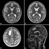
Case description: We report a 22-year-old woman admitted with headache, papilledema, vision loss, and a history of leukemia. Lumbar puncture revealed idiopathic intracranial hypertension (IIH). Three months after VPS implantation, she was readmitted with headache and worsening of visual impairment. CT evidenced a IPC with perilesional edema. Intraoperatively, a shunt revision and cyst drainage were opted for. We present a discussion and literature review on this unique complication of VPS, with emphasis on management.
Conclusion: It is important to understand and consider IPCs as complications of VPS surgery, including in adult patients and IIH cases.
Keywords: Catheter obstruction; Cerebrospinal fluid edema; Idiopathic intracranial hypertension; Intraparenchymal pericatheter cyst; Ventriculoperitoneal shunt.
Copyright: © 2022 Surgical Neurology International.
Conflict of interest statement
There are no conflicts of interest.
Figures


Similar articles
-
A Rare Case of Intraparenchymal Cerebrospinal Fluid Cyst Associated With Ventriculoperitoneal Shunt in an Adult Patient.Cureus. 2021 Aug 24;13(8):e17420. doi: 10.7759/cureus.17420. eCollection 2021 Aug. Cureus. 2021. PMID: 34589330 Free PMC article.
-
Intraparenchymal Pericatheter Cyst after Cerebrospinal Fluid Shunt: A Rare Complication with Challenging Diagnosis - Case Presentation and Review of the Literature.Asian J Neurosurg. 2019 Apr-Jun;14(2):581-584. doi: 10.4103/ajns.AJNS_288_18. Asian J Neurosurg. 2019. PMID: 31143289 Free PMC article.
-
Intraparenchymal pericatheter cyst. A rare complication of ventriculoperitoneal shunt for hydrocephalus.Br J Neurosurg. 2000 Jun;14(3):255-8. doi: 10.1080/026886900408487. Br J Neurosurg. 2000. PMID: 10912207
-
Review of the Management of Peroral Extrusion of Ventriculoperitoneal Shunt Catheter.J Clin Diagn Res. 2016 Nov;10(11):PE01-PE06. doi: 10.7860/JCDR/2016/23372.8920. Epub 2016 Nov 1. J Clin Diagn Res. 2016. PMID: 28050444 Free PMC article. Review.
-
A porencephalic cyst formation in a 6-year-old female with a functioning ventriculoperitoneal shunt: a case-based review.Childs Nerv Syst. 2018 Apr;34(4):611-616. doi: 10.1007/s00381-018-3725-x. Epub 2018 Jan 29. Childs Nerv Syst. 2018. PMID: 29380111 Review.
Cited by
-
Periventricular cyst as a complication of ventriculoperitoneal shunting in the context of intracranial haemorrhage: a case report and review of the literature.J Surg Case Rep. 2024 Jan 23;2024(1):rjad743. doi: 10.1093/jscr/rjad743. eCollection 2024 Jan. J Surg Case Rep. 2024. PMID: 38268536 Free PMC article.
References
-
- Aly B, Hammad W, Elkhouly H. Uncommon complications of ventriculoperitoneal shunt: Experience in 22 cases. Al-Azhar Assiut Med J. 2015;13:360–73.
-
- Balasubramaniam S, Tyagi DK, Sawant HV. Intraparenchymal pericatheter cyst following disconnection of ventriculoperitoneal shunt system. J Postgrad Med. 2013;59:232–4. - PubMed
-
- Bianchi F, Frassanito P, Tamburrini G, Caldarelli M, Massimi L. Shunt malfunction mimicking a cystic tumour. Br J Neurosurg. 2017;31:484–6. - PubMed
-
- Chiba Y, Takagi H, Nakajima F, Fujii S, Kitahara T, Yagishita S, et al. Cerebrospinal fluid edema: A rare complication of shunt operations for hydrocephalus. Report of three cases. J Neurosurg. 1982;57:697–700. - PubMed
Publication types
LinkOut - more resources
Full Text Sources
