. 2022 May 27:13:218.
doi: 10.25259/SNI_404_2022. eCollection 2022.

Anatomic variations of the floor of the third ventricle: Surgical implications for endoscopic third ventriculostomy

Affiliations

Anatomic variations of the floor of the third ventricle: Surgical implications for endoscopic third ventriculostomy

Nadin J Abdala-Vargas et al. Surg Neurol Int. .

Abstract

Background: Endoscopic third ventriculostomy (ETV) is currently used as a treatment for different types of hydrocephalus. However, the anatomical endoscopic variants of the third ventricle floor (3VF), as well as their surgical implications, have been underrated. The anatomic variations of the 3VF can influence the technique and the success rate of the ETV. The purpose of this article is to describe the anatomical variations of 3VF, assess their incidence, and discuss the implications for ETV.

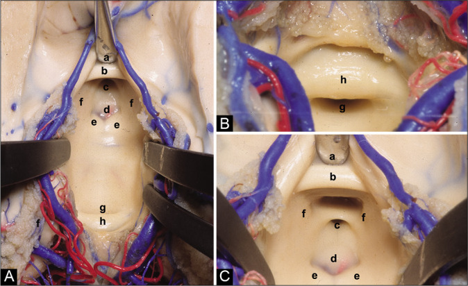
Methods: Intraoperative videos of 216 patients who underwent ETV between January 2012 and February 2020 at Hospital Infantil Universitario de San José, Bogotá, Colombia were reviewed. One hundred and eighty patients who met the criteria to demonstrate the type of 3VF were selected.

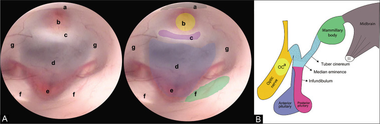
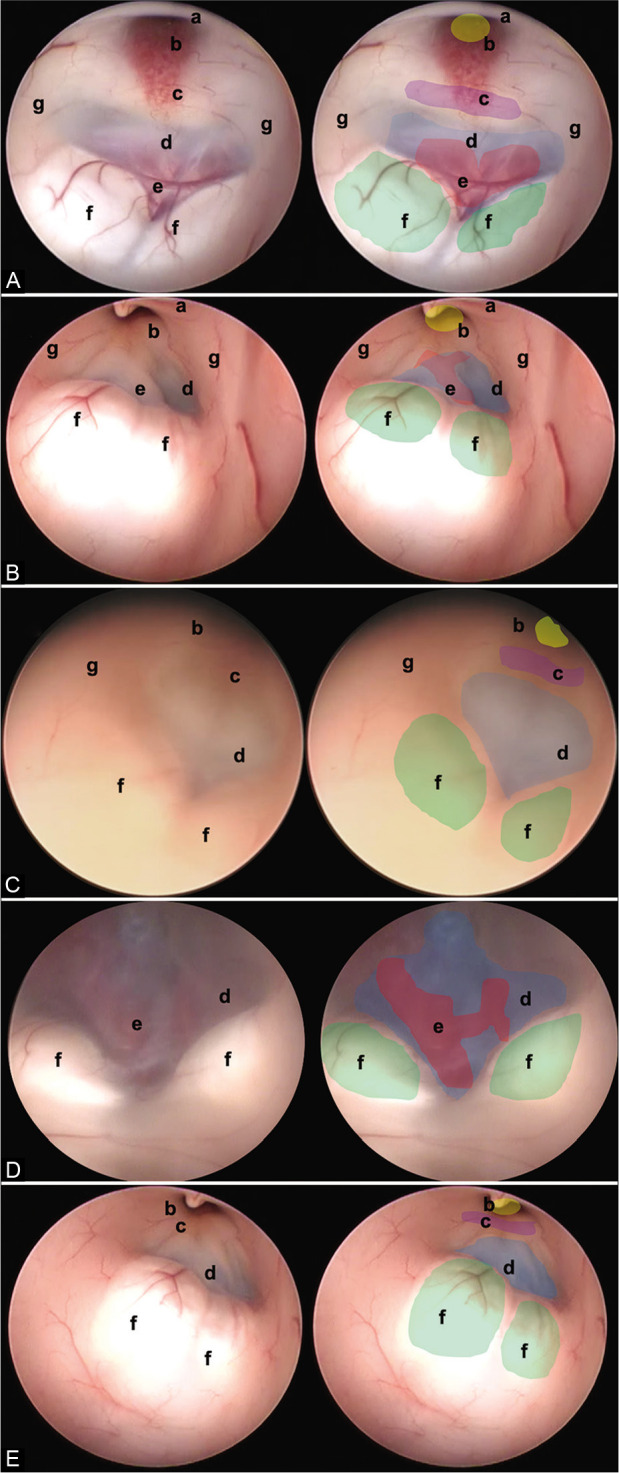
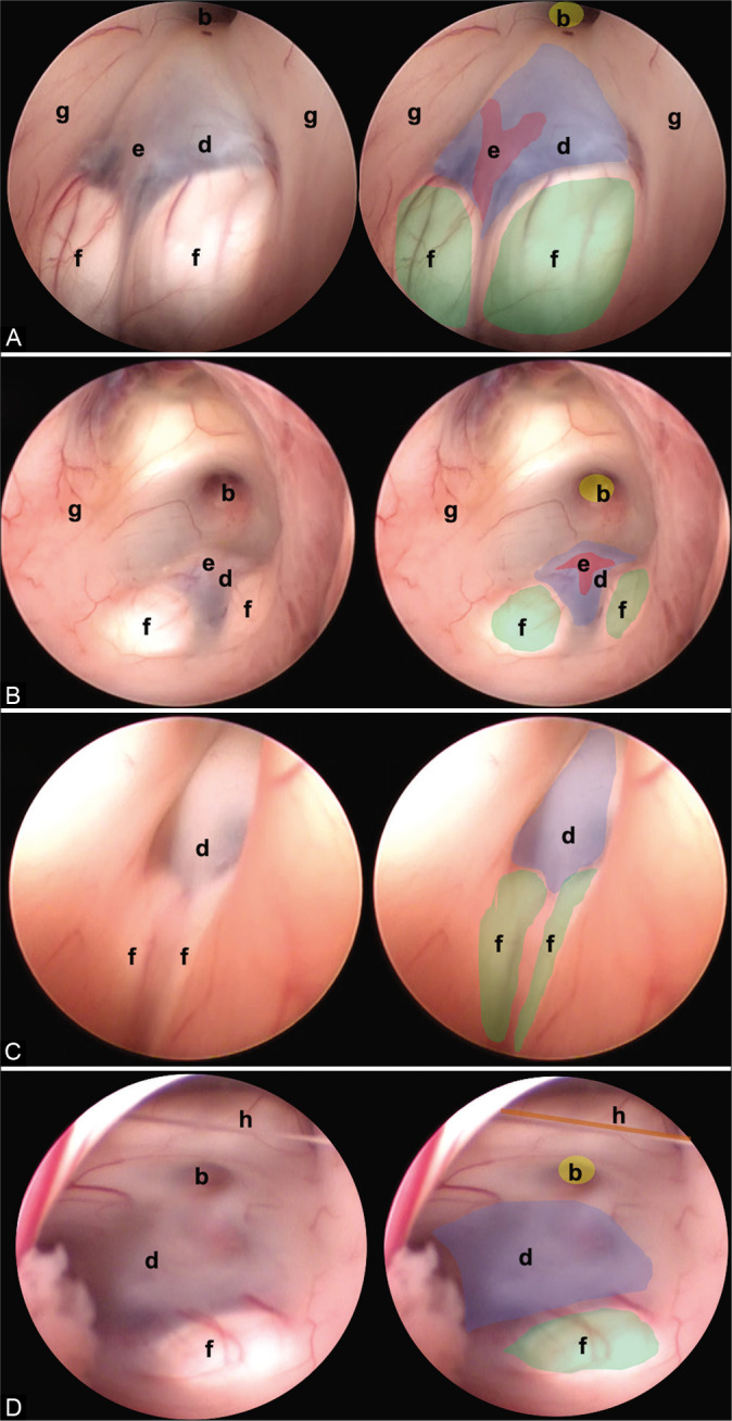
Results: 3VF types were classified as follows: (1) Thinned, (2) thickened, (3) partially erased, (4) globular or herniated, and (5) narrowed.

Conclusion: Knowledge of anatomical variations of the 3VF is paramount for ETV and it influences the success rate of the procedure.

Keywords: Cerebrospinal fluid shunt; Endoscopic third ventriculostomy; Hydrocephalus; Neuroendoscopy; Ventriculostomy.


Conflict of interest statement

There are no conflicts of interest.

Figures

Figure 1:
Figure 1:
(A) Endoscopic view of the FTV. In this image, the anatomical structures that make up the FTV are recognized, from anterior to posterior we find. (a) Optic chiasm, (b) infundibulum, (c) dorsum sellae, (d) floor of the third ventricle (tuber cinereum), (e) basilar complex, (f) mammillary bodies, and (g) hypothalamic wall. (B) Lateral view of the diencephalon. The structures that make up the floor of the third ventricle can be appreciated. (OCa) Optic chiasm.
Figure 2:
Figure 2:
(A) Anatomical superior view of the III ventricle floor. (B) Posterior superior view of the III ventricle floor. (C) Anterior superior view of the III ventricle floor. (a) Chiasmatic recess, (b) anterior commissure, (c) infundibular recess, (d) tuber cinereum, (e) mammillary bodies, (f) medial wall of the hypothalamus, (g) aqueduct, and (h) posterior commissure.
Figure 3:
Figure 3:
In this figure, it is possible to identify the different endoscopic anatomical variants of the FTV, in each image, a complete view of the ventricle is shown, and next to it is the representation of each of its structures. (A) Thinned floor, (B) thickened floor, (C) cleared floor, (D) globus or herniated floor, and (E) narrow floor. The anatomical structures are found as follows: (a) optic chiasm (black), (b) infundibulum (yellow), (c) dorsum sellae (purple), (d) floor of the third ventricle, (tuber cinereum) (blue), (e) basilar complex (red), (f) mammillary bodies (green), and (g) hypothalamic wall.
Figure 4:
Figure 4:
Other anatomic variants independent of the type of TVF can be identified. (A) Separate mammillary bodies, (B) the prominent basilar artery, (C) elongated third ventricle, and (D) interthalamic bands. The anatomical structures are found as follows: (b) infundibulum (yellow), (d) floor of the third ventricle (tuber cinereum) (blue), (e) basilar complex (red), (f) mammillary bodies (green), and (g) hypothalamic wall.
Figure 5:
Figure 5:
Two endoscopic anatomical variants are presented that we consider are not reported in the medical literature and that can constitute a surgical challenge for the surgeon in charge. (A) Translucent floor, (B) Y-floor. Anatomical structures are found as follows: (c) Dorsum sellae (purple), (d) floor of the third ventricle (tuber cinereum) (blue), (e) basilar complex (red), (f) mammillary bodies (green), (i) pituitary, and (j) brainstem.

References

    1. Anik I, Ceylan S, Koc K, Tugasaygi M, Sirin G, Gazioglu N, et al. Microsurgical and endoscopic anatomy of Liliequist’s membrane and the prepontine membranes: Cadaveric study and clinical implications. Acta Neurochir (Wien) 2011;153:1701–11. - PubMed
    1. Aydin S, Yilmazlar S, Aker S, Korfali E. Anatomy of the floor of the third ventricle in relation to endoscopic ventriculostomy. Clin Anat. 2009;22:916–24. - PubMed
    1. Corrales M, Torrealba G. The third ventricle. Normal anatomy and changes in some pathological conditions. Neuroradiology. 1976;11:271–7. - PubMed
    1. Demerdash A, Rocque BG, Johnston J, Rozzelle CJ, Yalcin B, Oskouian R, et al. Endoscopic third ventriculostomy: A historical review. Br J Neurosurg. 2017;31:28–32. - PMC - PubMed
    1. Dezena RA. 1st ed. Berlin, Germany: Springer International Publishing; 2017. Atlas of Endoscopic Neurosurgery of the Third Ventricle.

LinkOut - more resources