Tension pneumoventricle in a patient with a ventriculoperitoneal shunt and an ethmoidal meningoencephalocele
- PMID: 35673658
- PMCID: PMC9168403
- DOI: 10.25259/SNI_64_2022
Tension pneumoventricle in a patient with a ventriculoperitoneal shunt and an ethmoidal meningoencephalocele
Abstract
Background: Tension pneumoventricle is a rare, life-threatening complication. It has been rarely described in patients with ventriculoperitoneal (VP) shunts.
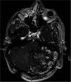
Case description: A 28-year-old male patient with a VP shunt became progressively lethargic after falling from his wheelchair. Skull X-rays and head CT scan showed abundant air inside the ventricles. He was taken to the operating room, and the shunt was revised without improvement. Two days later, a frontal external ventricular drain was placed to remove the air. In the investigation toward the etiology of the pneumoventricle, a review of previous head CT scans and brain MRIs showed that the patient had a small left frontonasal meningoencephalocele extending into the ethmoid, which had been unnoticed. He underwent repair of the defect with adequate sealing of the frontal skull base.
Conclusion: In a shunted patient with moderate or severe symptoms from a tension pneumoventricle, external ventricular drainage is required to remove the air as the shunt is inadequate.
Keywords: Ethmoid; Meningoencephalocele; Pneumoventricle; Tension; Ventriculoperitoneal shunt.
Copyright: © 2022 Surgical Neurology International.
Conflict of interest statement
There are no conflicts of interest.
Figures


Similar articles
-
Tension pneumoventricle: Reversible cause for aphasia.Qatar Med J. 2021 Apr 23;2021(1):15. doi: 10.5339/qmj.2021.15. eCollection 2021. Qatar Med J. 2021. PMID: 33959489 Free PMC article.
-
Pathogenesis of Delayed Tension Intraventricular Pneumocephalus in Shunted Patient: Possible Role of Nocturnal Positive Pressure Ventilation.World Neurosurg. 2016 Jan;85:365.e17-20. doi: 10.1016/j.wneu.2015.09.001. Epub 2015 Sep 9. World Neurosurg. 2016. PMID: 26363220
-
Tension Pneumoventricle After Endoscopic Transsphenoidal Surgery for Rathke Cleft Cyst.World Neurosurg. 2020 Mar;135:228-232. doi: 10.1016/j.wneu.2019.12.065. Epub 2019 Dec 19. World Neurosurg. 2020. PMID: 31863895
-
Quadriparesis Due to Delayed Tension Pneumoventricle.Neurologist. 2021 Nov 26;27(2):74-78. doi: 10.1097/NRL.0000000000000363. Neurologist. 2021. PMID: 34842575 Review.
-
Tension Pneumoventricle Secondary to Cutaneous-Ventricular Fistula: Case Report and Literature Review.World Neurosurg. 2020 Oct;142:155-158. doi: 10.1016/j.wneu.2020.06.145. Epub 2020 Jun 26. World Neurosurg. 2020. PMID: 32599189 Review.
Cited by
-
Tension pneumocephalus in a patient with NF1 following ventriculoperitoneal shunt-deciphering the cause and proposed management strategy.Childs Nerv Syst. 2023 Dec;39(12):3601-3606. doi: 10.1007/s00381-023-06052-6. Epub 2023 Jul 1. Childs Nerv Syst. 2023. PMID: 37392224 Review.
References
-
- Almubarak AO, Fakhroo F, Alhuthayl MR, Kanaan I, Aldahash H. Tension pneumoventricle secondary to cutaneous-ventricular fistula: Case report and literature review. World Neurosurg. 2020;142:155–8. - PubMed
-
- Ani CC, Ismaila BO. Tension pneumoventricle: A report of two cases. Niger J Clin Pract. 2016;19:559–62. - PubMed
-
- Armocida D, Pesce A, Frati A, Miscusi M, Paglia F, Raco A. Pneumoventricle of unknown origin: A personal experience and literature review of a clinical enigma. World Neurosurg. 2019;122:661–4. - PubMed
-
- Cartwright MJ, Eisenberg MB. Tension pneumocephalus associated with rupture of a middle fossa encephalocele. Case report. J Neurosurg. 1992;76:292–5. - PubMed
-
- Chhiber SS, Nizami FA, Kirmani AR, Wani MA, Bhat AR, Zargar J, et al. Delayed posttraumatic intraventricular tension pneumocephalus: Case report. Neurosurg Q. 2011;21:128–32.
Publication types
LinkOut - more resources
Full Text Sources
