

Cervical cytology: Radiation and other therapy effects
- PMID: 35673693
- PMCID: PMC9168396
- DOI: 10.25259/CMAS_03_12_2021
Cervical cytology: Radiation and other therapy effects
Abstract
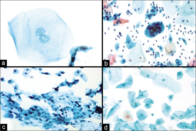
The different treatment options for carcinoma cervix include radiation, chemotherapy, and surgical treatments. Cytological analysis of smears is crucial for patient follow-up to determine response to therapy and to diagnose the persistence or recurrence of malignancy. Anatomical alterations and changes in cell morphology following radiation or chemotherapy make collecting and interpreting cervical cytology samples difficult. These issues can be mitigated by liquid-based cytology. Ionizing radiation is used in radiotherapy (RT) to kill cells. It is important that cytologists are aware of alterations in morphology of the cells. Radiation can cause cytoplasmic and nuclear changes. Cellular enlargement, vacuolation, granularity loss, and other changes linked with cell death are examples of cytoplasmic alterations. Nuclear enlargement and multinucleation are the most frequent nuclear alterations. These changes are determined by the amount of time that has passed since radiation. It should be emphasized that no one characteristic is pathognomonic. Post-irradiation dysplasia is a condition described as abnormal cellular changes in non-neoplastic epithelial cells after RT. Chemotherapy causes comparable alterations as radiation but impacts fewer cells. Busulfan and other chemotherapeutic treatments may produce morphological alterations, which cytologists must be aware of and able to identify. Immunosuppressive treatments, hormonal therapy, and tamoxifen are some of the other drugs that might cause changes in cervical morphology. Surgical methods used in the detection and treatment of cervical cancer may potentially cause alterations as a result of thermal damage and healing. For the treatment of cervical lesions, electrocautery and the loop electrosurgical excisional procedure are available. These procedures employ electric current ablation leading to ischemic changes in the cervical smear. Cytological analysis of smears following treatment with these modalities necessitates a comprehensive history, kind of therapy, and duration of treatment.
Keywords: Cervical Smear; Cytology; Pap smear; Radiation; Uterine Cervical Neoplasms.
© 2022 Cytopathology Foundation Inc, Published by Scientific Scholar.
Figures


Similar articles
-
[Health technology assessment report: Computer-assisted Pap test for cervical cancer screening].Epidemiol Prev. 2012 Sep-Oct;36(5 Suppl 3):e1-43. Epidemiol Prev. 2012. PMID: 23139174 Italian.
-
Cervical Papanicolaou Smears in Hematopoietic Stem Cell Transplant Recipients: High Prevalence of Therapy-Related Atypia during the Acute Phase.Biol Blood Marrow Transplant. 2017 Aug;23(8):1367-1373. doi: 10.1016/j.bbmt.2017.04.022. Epub 2017 Apr 24. Biol Blood Marrow Transplant. 2017. PMID: 28450182 Clinical Trial.
-
Post-irradiation cytology of cervical cancer patients.Cytopathology. 1992;3(3):167-82. doi: 10.1111/j.1365-2303.1992.tb00043.x. Cytopathology. 1992. PMID: 1511122 Review.
-
[A falsely reassuring cervical smear in adenocarcinoma of the external os].Ned Tijdschr Geneeskd. 2008 Apr 26;152(17):977-80. Ned Tijdschr Geneeskd. 2008. PMID: 18549169 Dutch.
-
Cytopathologic evaluation of patients submitted to radiotherapy for uterine cervix cancer.Rev Assoc Med Bras (1992). 2017 Apr;63(4):379-385. doi: 10.1590/1806-9282.63.04.379. Rev Assoc Med Bras (1992). 2017. PMID: 28614543 Review.
Cited by
-
HSP90 regulates dCK stability and inhibits ionizing radiation-induced ferroptosis in cervical cancer cells.Cell Death Discov. 2025 Apr 22;11(1):191. doi: 10.1038/s41420-025-02388-x. Cell Death Discov. 2025. PMID: 40263268 Free PMC article.
-
Clinical trial comparing the use of Orcellex® Brush versus Cervex-Brush® on vaginal vault smear cytology adequacy rate in patients treated with radiotherapy for cervical cancer.J Gynecol Oncol. 2025 May;36(3):e43. doi: 10.3802/jgo.2025.36.e43. Epub 2024 Oct 22. J Gynecol Oncol. 2025. PMID: 39453394 Free PMC article. Clinical Trial.
References
-
- Rana MK, Singh K, Mahajan MK, Singh Rana AP. Clinicopathological profile of cervical carcinoma: An experience of tertiary care cancer centre. Asian Pac J Cancer Care. 2019;4:83–6.
-
- Padilha CM, Araújo ML, de Souza SA. Cytopathologic evaluation of patients submitted to radiotherapy for uterine cervix cancer. Rev Assoc Medica Bras 1992. 2017;63:379–85. - PubMed
Publication types
LinkOut - more resources
Full Text Sources
Research Materials