

Postoperative retinal microstructure and functional outcome after inverted-flap technique associated with silicone oil tamponade in macular hole surgery
- PMID: 35673818
- PMCID: PMC9289692
- DOI: 10.47162/RJME.62.4.11
Postoperative retinal microstructure and functional outcome after inverted-flap technique associated with silicone oil tamponade in macular hole surgery
Abstract
Purpose: Our retrospective study on 27 patients with a large mean macular hole diameter (MH-D) of 480.08±78.62 μm evaluates the usefulness of combining the current internal limiting membrane (ILM) inverted-flap surgical technique with silicone oil tamponade, which has been associated with the classical technique of ILM peeling.
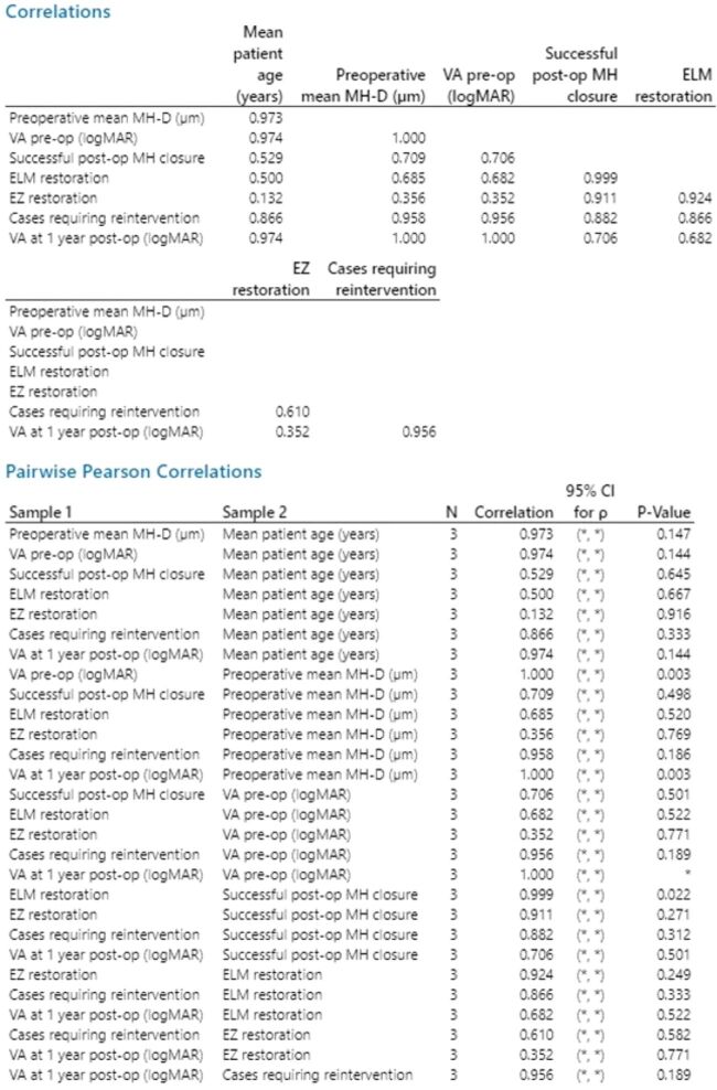
Results: Functional results: mean visual acuity (VA) improved to 0.89±0.11 logMar (logarithm of the minimum angle of resolution, at one month), 0.67±0.03 logMar (at three months), 0.52±0.04 logMar (at six months), 0.42±0.15 logMar (at one year) postoperative (final VA), with statistical linkage between preoperative VA and final VA (two-sample t-test, p=0.007), mean MH-D and final VA (regression analysis, p=0.003). We compared the results by MH size (Group A ≤400 μm - eight eyes and Group B >400 μm - 19 eyes), finding statistical variance (Bonett & Levene methods). Group A presented a final VA of 0.21±0.12 logMar, while Group B had 0.51±0.17 logMar. Successful closure was noted in 25 (92.59%) cases, with Group A having complete closure and external limiting membrane (ELM) restoration with ellipsoid zone (EZ) regeneration in six cases. Group B had successful closure in 17 (89.47%) cases with ELM restoration in 16 cases and EZ regeneration in seven (38.88%) cases, with reintervention in two cases. Restoration of the ELM was correlated [Pearson's correlation coefficient (PCC) of 0.999, p=0.022] with successful closure, with overall restoration obtained in 24 (88.88%) cases and EZ regeneration in 13 (48.14%) cases.
Conclusions: ILM inverted-flap technique with silicone oil tamponade had favorable functional and anatomical outcomes. ELM restoration was associated with successful MH closure.
Conflict of interest statement
The authors declare that they have no competing interests or conflict of interests.
Figures


Similar articles
-
EFFECT OF INVERTED INTERNAL LIMITING MEMBRANE FLAP ON CLOSURE RATE, POSTOPERATIVE VISUAL ACUITY, AND RESTORATION OF OUTER RETINAL LAYERS IN PRIMARY IDIOPATHIC MACULAR HOLE SURGERY.Retina. 2020 Oct;40(10):1955-1963. doi: 10.1097/IAE.0000000000002707. Retina. 2020. PMID: 31834129
-
Inverted internal limiting membrane flap technique versus complete internal limiting membrane peeling in large macular hole surgery: a comparative study.BMC Ophthalmol. 2020 Jan 6;20(1):11. doi: 10.1186/s12886-019-1294-8. BMC Ophthalmol. 2020. PMID: 31907015 Free PMC article.
-
Large Idiopathic Macular Hole Surgery: Remodelling of Outer Retinal Layers after Traditional Internal Limiting Membrane Peeling or Inverted Flap Technique.Ophthalmologica. 2020;243(5):334-341. doi: 10.1159/000505926. Epub 2020 Jan 15. Ophthalmologica. 2020. PMID: 31940651
-
Comparative efficacy evaluation of inverted internal limiting membrane flap technique and internal limiting membrane peeling in large macular holes: a systematic review and meta-analysis.BMC Ophthalmol. 2020 Jan 8;20(1):14. doi: 10.1186/s12886-019-1271-2. BMC Ophthalmol. 2020. PMID: 31914954 Free PMC article.
-
Inverted Internal Limiting Membrane Flap versus Internal Limiting Membrane Peeling for <400 μm Macular Hole: A Meta-Analysis and Systematic Review.Ophthalmic Res. 2023;66(1):1342-1352. doi: 10.1159/000534873. Epub 2023 Nov 6. Ophthalmic Res. 2023. PMID: 37931613 Free PMC article.
References
-
- Duker JS, Kaiser PK, Binder S, de Smet MD, Gaudric A, Reichel E, Sadda SR, Sebag J, Spaide RF, Stalmans P. The International Vitreomacular Traction Study Group classification of vitreomacular adhesion, traction, and macular hole. Ophthalmology. 2013;120(12):2611–2619. - PubMed
-
- Kalur A, Muste J, Singh RP. A review of surgical techniques for the treatment of large idiopathic macular holes. Ophthalmic Surg Lasers Imaging Retina. 2022;53(1):52–61. - PubMed
-
- Schneider EW, Jaffe GJ. Baseline characteristics of vitreomacular traction progressing to full-thickness macular or lamellar holes in the phase III trials of enzymatic vitreolysis. Retina. 2020;40(8):1579–1584. - PubMed
-
- Oh J, Smiddy WE, Flynn HW, Gregori G, Lujan B. Photoreceptor inner/outer segment defect imaging by spectral domain OCT and visual prognosis after macular hole surgery. Invest Ophthalmol Vis Sci. 2010;51(3):1651–1658. - PubMed
MeSH terms
Substances
LinkOut - more resources
Full Text Sources
