

Esophageal spindle cell lipoma
- PMID: 35673825
- PMCID: PMC9289690
- DOI: 10.47162/RJME.62.4.18
Esophageal spindle cell lipoma
Abstract
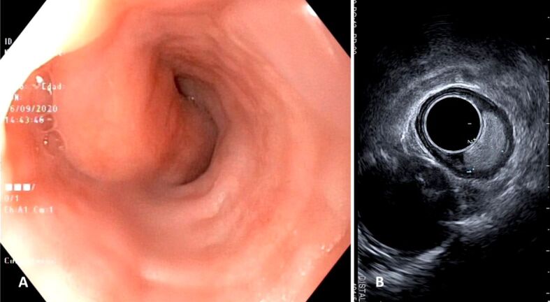
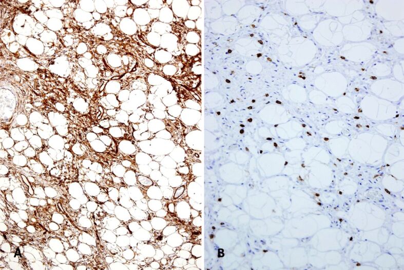
Symptomatic ordinary esophageal lipomas are rare tumors. Spindle cell lipomas (SCLs) of this location are even more infrequent. To our knowledge, only a previous esophageal SCL case has been reported. We describe herein the case of a 62-year-old woman with a long history of heartburn and feeling of abdominal distension. Preoperative investigations, including a Barium meal, gastroscopy, and echoendoscopy revealed a lipomatous polypoid mass attached to the middle esophageal segment. The lesion (3.5×2×1 cm) was excised endoscopically under deep sedation. The final histopathology diagnosis was pedunculated SCL. An accurate diagnosis of esophageal SCL is crucial to rule out malignant lesions, relieve symptoms, and undertake suitable treatment. The main differential diagnosis includes well-differentiated sclerosing liposarcoma, atypical spindle cell∕pleomorphic lipomatous tumor, giant fibrovascular polyp, and fat-forming solitary fibrous tumor. Although rare, SCL should be added to the list of lipomatous tumors that can affect the esophagus. Complete excision is the appropriate treatment.
Conflict of interest statement
The authors declare that they have no conflict of interests.
Figures


Similar articles
-
Polypoid fibroadipose tumors of the esophagus: 'giant fibrovascular polyp' or liposarcoma? A clinicopathological and molecular cytogenetic study of 13 cases.Mod Pathol. 2018 Feb;31(2):337-342. doi: 10.1038/modpathol.2017.140. Epub 2017 Oct 6. Mod Pathol. 2018. PMID: 28984298
-
Liposarcoma arising in a giant lipomatous polyp of the esophagus.Korean J Intern Med. 1989 Jan;4(1):86-9. doi: 10.3904/kjim.1989.4.1.86. Korean J Intern Med. 1989. PMID: 2487410 Free PMC article.
-
FDG PET/CT findings in a rare case of giant fibrovascular polyp of the esophagus harboring atypical lipomatous tumor/well-differentiated liposarcoma.Clin Nucl Med. 2014 Mar;39(3):288-91. doi: 10.1097/RLU.0000000000000358. Clin Nucl Med. 2014. PMID: 24458178
-
Lipomatous tumors.Monogr Pathol. 1996;38:207-39. Monogr Pathol. 1996. PMID: 8744279 Review.
-
Atypical lipomatous tumor mimicking giant fibrovascular polyp of the esophagus: report of a case and a critical review of literature.Hum Pathol. 2013 Jun;44(6):1165-70. doi: 10.1016/j.humpath.2012.10.023. Epub 2013 Jan 24. Hum Pathol. 2013. PMID: 23352209 Review.
Cited by
-
Giant spindle cell lipoma of the left inguinal region: A rare case with diagnostic challenges on MRI.Radiol Case Rep. 2025 Jun 10;20(9):4262-4265. doi: 10.1016/j.radcr.2025.05.034. eCollection 2025 Sep. Radiol Case Rep. 2025. PMID: 40547940 Free PMC article.
References
-
- Tang LH, Lao QY, Yu L, Wang J. Spindle cell lipoma and pleomorphic lipoma: a clinicopathologic analysis of 65 cases] Zhonghua Bing Li Xue Za Zhi. 2018;47(4):263–268. - PubMed
-
- Ud Din N, Zhang P, Sukov WR, Sattler CA, Jenkins SM, Doyle LA, Folpe AL, Fritchie KJ. Spindle cell lipomas arising at atypical locations. Am J Clin Pathol. 2016;146(4):487–495. - PubMed
-
- Razzak R, Bédard ELR, Hunt I, Satkunam N. Spindle cell lipoma of the esophagus. Eur J Cardiothorac Surg. 2009;35(3):542–543. - PubMed
Publication types
MeSH terms
LinkOut - more resources
Full Text Sources
Medical
Research Materials
Miscellaneous
