

Computational lung modelling in respiratory medicine
- PMID: 35673857
- PMCID: PMC9174712
- DOI: 10.1098/rsif.2022.0062
Computational lung modelling in respiratory medicine
Abstract
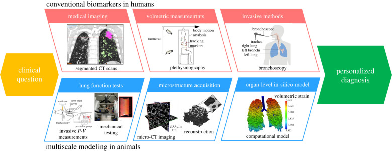
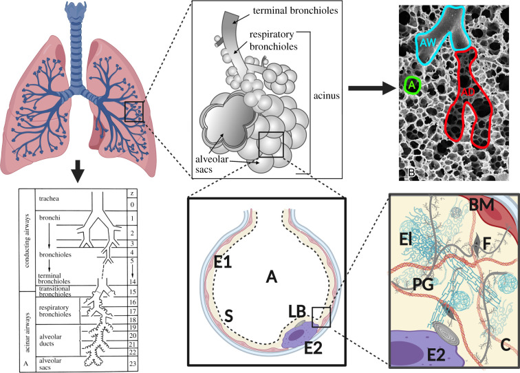
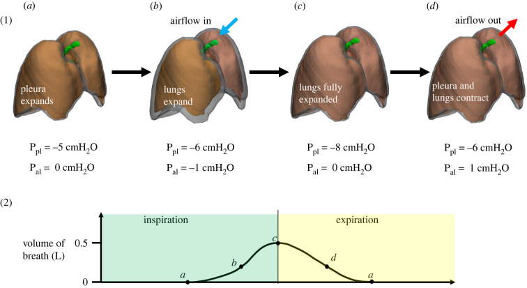
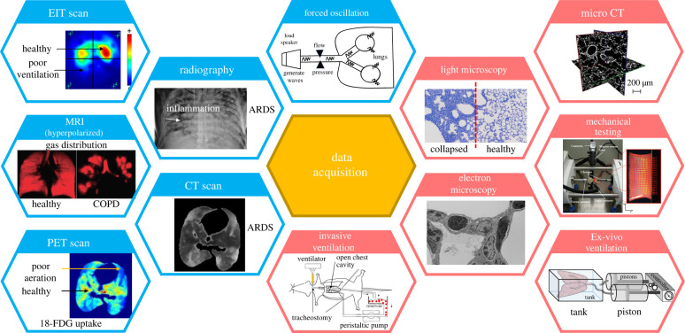
Computational modelling of the lungs is an active field of study that integrates computational advances with lung biophysics, biomechanics, physiology and medical imaging to promote individualized diagnosis, prognosis and therapy evaluation in lung diseases. The complex and hierarchical architecture of the lung offers a rich, but also challenging, research area demanding a cross-scale understanding of lung mechanics and advanced computational tools to effectively model lung biomechanics in both health and disease. Various approaches have been proposed to study different aspects of respiration, ranging from compartmental to discrete micromechanical and continuum representations of the lungs. This article reviews several developments in computational lung modelling and how they are integrated with preclinical and clinical data. We begin with a description of lung anatomy and how different tissue components across multiple length scales affect lung mechanics at the organ level. We then review common physiological and imaging data acquisition methods used to inform modelling efforts. Building on these reviews, we next present a selection of model-based paradigms that integrate data acquisitions with modelling to understand, simulate and predict lung dynamics in health and disease. Finally, we highlight possible future directions where computational modelling can improve our understanding of the structure-function relationship in the lung.
Keywords: computational modelling; lung biomechanics; lung biophysical models; lung imaging.
Figures
Similar articles
-
Short-Term Memory Impairment.2024 Jun 8. In: StatPearls [Internet]. Treasure Island (FL): StatPearls Publishing; 2025 Jan–. 2024 Jun 8. In: StatPearls [Internet]. Treasure Island (FL): StatPearls Publishing; 2025 Jan–. PMID: 31424720 Free Books & Documents.
-
Measures implemented in the school setting to contain the COVID-19 pandemic.Cochrane Database Syst Rev. 2022 Jan 17;1(1):CD015029. doi: 10.1002/14651858.CD015029. Cochrane Database Syst Rev. 2022. Update in: Cochrane Database Syst Rev. 2024 May 2;5:CD015029. doi: 10.1002/14651858.CD015029.pub2. PMID: 35037252 Free PMC article. Updated.
-
Survivor, family and professional experiences of psychosocial interventions for sexual abuse and violence: a qualitative evidence synthesis.Cochrane Database Syst Rev. 2022 Oct 4;10(10):CD013648. doi: 10.1002/14651858.CD013648.pub2. Cochrane Database Syst Rev. 2022. PMID: 36194890 Free PMC article.
-
Assessing the comparative effects of interventions in COPD: a tutorial on network meta-analysis for clinicians.Respir Res. 2024 Dec 21;25(1):438. doi: 10.1186/s12931-024-03056-x. Respir Res. 2024. PMID: 39709425 Free PMC article. Review.
-
Comparison of Two Modern Survival Prediction Tools, SORG-MLA and METSSS, in Patients With Symptomatic Long-bone Metastases Who Underwent Local Treatment With Surgery Followed by Radiotherapy and With Radiotherapy Alone.Clin Orthop Relat Res. 2024 Dec 1;482(12):2193-2208. doi: 10.1097/CORR.0000000000003185. Epub 2024 Jul 23. Clin Orthop Relat Res. 2024. PMID: 39051924
Cited by
-
Asymmetric lung increases particle filtration by deposition.Sci Rep. 2023 Jun 3;13(1):9040. doi: 10.1038/s41598-023-36176-3. Sci Rep. 2023. PMID: 37270569 Free PMC article.
-
Stress-strain curve and elastic behavior of the fibrotic lung with usual interstitial pneumonia pattern during protective mechanical ventilation.Sci Rep. 2024 Jun 7;14(1):13158. doi: 10.1038/s41598-024-63670-z. Sci Rep. 2024. PMID: 38849437 Free PMC article.
-
A new computational framework for simulating airway resistance, fraction of exhaled nitric oxide, and diffusing capacity for nitric oxide.PLoS One. 2025 Jan 30;20(1):e0311667. doi: 10.1371/journal.pone.0311667. eCollection 2025. PLoS One. 2025. PMID: 39883668 Free PMC article.
-
Towards constructing a generalized structural 3D breathing human lung model based on experimental volumes, pressures, and strains.PLoS Comput Biol. 2025 Jan 13;21(1):e1012680. doi: 10.1371/journal.pcbi.1012680. eCollection 2025 Jan. PLoS Comput Biol. 2025. PMID: 39804822 Free PMC article.
-
Physics-informed motion registration of lung parenchyma across static CT images.ArXiv [Preprint]. 2024 Jul 3:arXiv:2407.03457v1. ArXiv. 2024. Update in: Annu Int Conf IEEE Eng Med Biol Soc. 2024 Jul;2024:1-4. doi: 10.1109/EMBC53108.2024.10781530. PMID: 39010873 Free PMC article. Updated. Preprint.
References
Publication types
MeSH terms
Grants and funding
LinkOut - more resources
Full Text Sources
