

[P4HA2 promotes occurrence and progression of liver cancer by regulating the PI3K/Akt/mTOR signaling pathway]
- PMID: 35673909
- PMCID: PMC9178641
- DOI: 10.12122/j.issn.1673-4254.2022.05.06
[P4HA2 promotes occurrence and progression of liver cancer by regulating the PI3K/Akt/mTOR signaling pathway]
Abstract
Objective: To investigate the role of proline 4-hydroxylase Ⅱ (P4HA2) in the occurrence and progression of liver cancer.
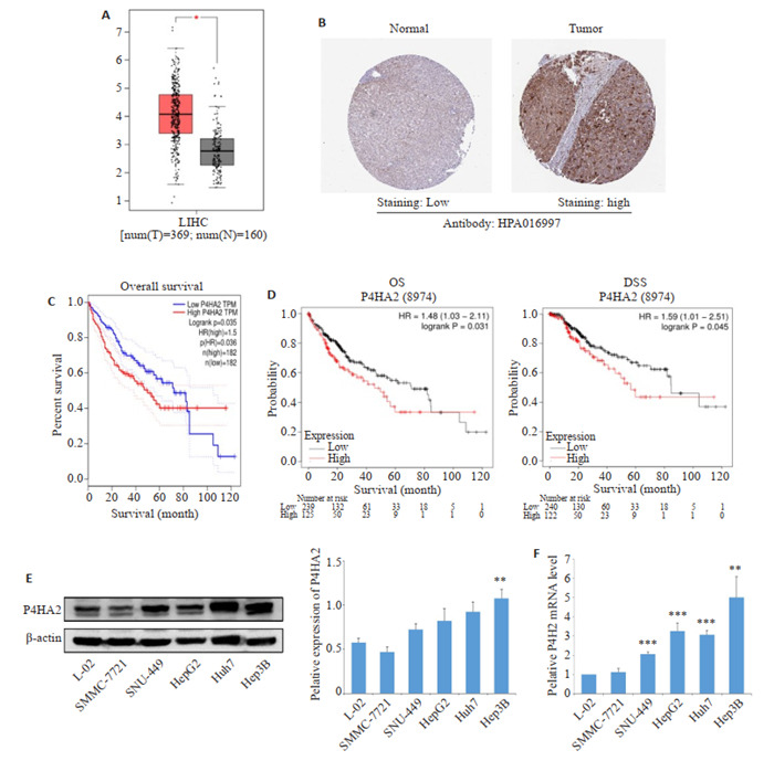
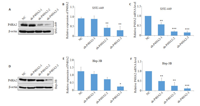
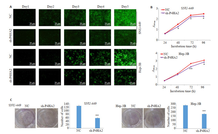
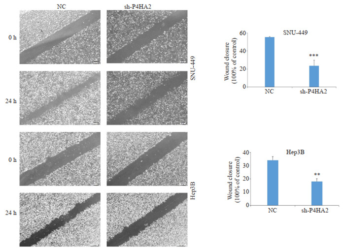
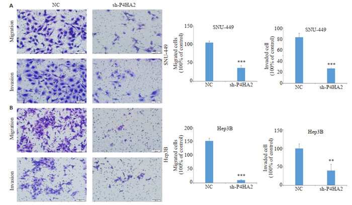
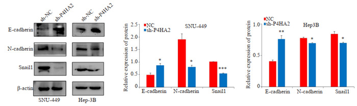
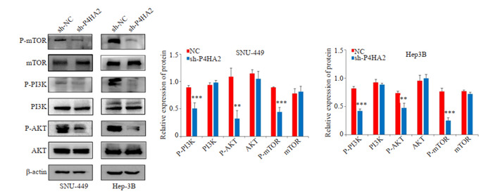
Methods: GEPIA and Human Protein Atlas database were used to predict the expression of P4HA2 in hepatocellular carcinoma (HCC), and K-M plotter online database was used to analyze the relationship between P4HA2 expression and the prognosis of HCC. We also examined the expressions of P4HA2 in HCC cells and normal hepatocytes using qRT-PCR and Western blotting. With lentivirus-mediated RNA interference, P4HA2 expression was knocked down in hepatoma SNU-449 and Hep-3B cells, and the changes in cell proliferation, migration and invasion were assessed using cell counting kit-8 (CCK-8) assay, colony formation test, scratch test and Transwell assay. The changes in the expressions of epithelial-mesenchymal transition (EMT) and PI3K/Akt/mTOR signal pathway-related proteins were detected using Western blotting.
Results: Online database analysis showed that the expression of P4HA2 was significantly higher in HCC tissues than in normal liver tissues (P < 0.05). The expression levels of P4HA2 mRNA and protein were also significantly higher in HCC cell lines than in normal hepatocytes (P < 0.01). Lentivirus-mediated RNA interference of P4HA2 significantly lowered the expression levels of P4HA2 mRNA and protein in the hepatoma cells (P < 0.05) and caused obvious inhibition of cell proliferation, migration and invasion. P4HA2 knockdown significantly increased the expression of E-cadherin protein, lowered the expressions of N-cadherin and Snail, and obviously decreased the expressions of phosphorylated PI3K, AKT and mTOR (P < 0.05).
Conclusion: P4HA2 enhances the proliferation, migration, invasion, and EMT of hepatoma cells by activating the PI3K/Akt/mTOR signaling pathway to promote the occurrence and progression of liver cancer.
目的: 探讨脯氨酸4-羟化酶Ⅱ(P4HA2)在肝癌细胞发生发展中的作用及相关机制。
方法: 利用GEPIA、Human Protein Atlas数据库预测P4HA2在肝癌中的表达情况,利用K-M plotter在线数据库分析P4HA2的表达情况与肝癌预后的关系,采用qRT-PCR和Western blot检测肝癌细胞和正常肝细胞中P4HA2的表达。构建慢病毒载体,用携带P4HA2 shRNA和Con shRNA的慢病毒载体分别转染肝癌SNU-449和Hep-3B细胞系,建立沉默表达P4HA2的细胞株(shP4HA2组)和对照组细胞株(NC组)。采用CCK-8、集落形成试验、划痕实验和Transwell实验分别检测细胞增殖、迁移和侵袭能力。采用Western blot实验检测上皮-间质转化和PI3K/Akt/mTOR信号通路相关蛋白表达情况。
结果: 在线数据库分析结果显示,肝癌组织中P4HA2表达高于正常肝组织(P < 0.05)。同时,肝癌细胞系中P4HA2 mRNA和蛋白表达水平也高于正常肝细胞(P < 0.01)。慢病毒干扰后,与NC组相比,shP4HA2组中mRNA和蛋白表达水平下降(P < 0.05)。P4HA2基因表达沉默后,细胞的增殖、迁移和侵袭受到抑制。Western blot显示,相对于NC组,shP4HA2组的E-cadherin蛋白表达上升,N-cadherin、Snail蛋白表达下降(P < 0.05),在PI3K/ AKT/mTOR通路中,磷酸化的PI3K(P-PI3K)、AKT(P-AKT)和mTOR(P-mTOR)显示出较低的水平(P < 0.05)。
结论: P4HA2通过激活PI3K/Akt/mTOR信号通路影响肝癌细胞的增殖、迁移、侵袭,促进肝癌的发生发展。
Keywords: PI3K/AKT/mTOR pathway; hepatocellular carcinoma; invasion and migration; prolyl-4-hydroxylase alpha polypeptide Ⅱ.
Figures
References
-
- Allemani C, Weir HK, Carreira H, et al. Global surveillance of cancer survival 1995-2009: analysis of individual data for 25, 676, 887 patients from 279 population-based registries in 67 countries (CONCORD-2. Lancet. 2015;385(9972):977–1010. doi: 10.1016/S0140-6736(14)62038-9. [Allemani C, Weir HK, Carreira H, et al. Global surveillance of cancer survival 1995-2009: analysis of individual data for 25, 676, 887 patients from 279 population-based registries in 67 countries (CONCORD-2[) J]. Lancet, 2015, 385(9972): 977-1010.] - DOI - PMC - PubMed
-
- Serper M, Taddei TH, Mehta R, et al. Association of provider specialty and multidisciplinary care with hepatocellular carcinoma treatment and mortality. Gastroenterology. 2017;152(8):1954–64. doi: 10.1053/j.gastro.2017.02.040. [Serper M, Taddei TH, Mehta R, et al. Association of provider specialty and multidisciplinary care with hepatocellular carcinoma treatment and mortality[J]. Gastroenterology, 2017, 152(8): 1954-64.] - DOI - PMC - PubMed
-
- Zheng XL, Liu WY, Xiang JX, et al. Collagen I promotes hepatocellular carcinoma cell proliferation by regulating integrin β1/ FAK signaling pathway in nonalcoholic fatty liver. Oncotarget. 2017;8(56):95586–95. doi: 10.18632/oncotarget.21525. [Zheng XL, Liu WY, Xiang JX, et al. Collagen I promotes hepatocellular carcinoma cell proliferation by regulating integrin β1/ FAK signaling pathway in nonalcoholic fatty liver[J]. Oncotarget, 2017, 8(56): 95586-95.] - DOI - PMC - PubMed
MeSH terms
Substances
LinkOut - more resources
Full Text Sources
Medical
Research Materials
Miscellaneous