Review
. 2022 Aug;111(3):662-682.
doi: 10.1111/tpj.15859. Epub 2022 Jun 27.

B vitamin supply in plants and humans: the importance of vitamer homeostasis

Affiliations
Review

B vitamin supply in plants and humans: the importance of vitamer homeostasis

Zeguang Liu et al. Plant J. 2022 Aug.

Abstract

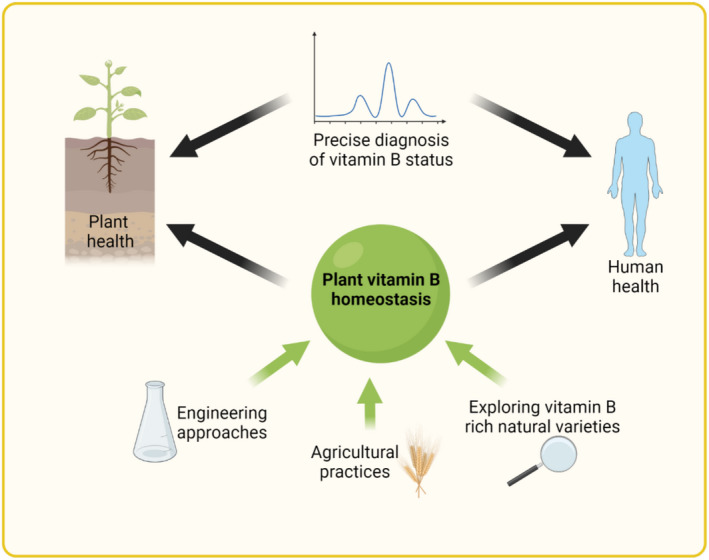
B vitamins are a group of water-soluble micronutrients that are required in all life forms. With the lack of biosynthetic pathways, humans depend on dietary uptake of these compounds, either directly or indirectly, from plant sources. B vitamins are frequently given little consideration beyond their role as enzyme accessory factors and are assumed not to limit metabolism. However, it should be recognized that each individual B vitamin is a family of compounds (vitamers), the regulation of which has dedicated pathways. Moreover, it is becoming increasingly evident that individual family members have physiological relevance and should not be sidelined. Here, we elaborate on the known forms of vitamins B1 , B6 and B9 , their distinct functions and importance to metabolism, in both human and plant health, and highlight the relevance of vitamer homeostasis. Research on B vitamin metabolism over the past several years indicates that not only the total level of vitamins but also the oft-neglected homeostasis of the various vitamers of each B vitamin is essential to human and plant health. We briefly discuss the potential of plant biology studies in supporting human health regarding these B vitamins as essential micronutrients. Based on the findings of the past few years we conclude that research should focus on the significance of vitamer homeostasis - at the organ, tissue and subcellular levels - which could improve the health of not only humans but also plants, benefiting from cross-disciplinary approaches and novel technologies.

Keywords: B vitamins; coenzymes; homeostasis; human health; plant health; vitamer.


Conflict of interest statement

The authors declare that they have no conflicts of interest associated with this work.

Figures

Figure 1
Figure 1
Vitamin B1 metabolism in plants and humans. (a) Generalized chemical structure of vitamin B1. The basic unit thiamine (hashed box) consists of a pyrimidine (green) and thiazole (blue) heterocycle, bridged by a methylene group (yellow). Thiamine derivatives vary in phosphorylation states (black, n = 1–3), as thiamine monophosphate (TMP), thiamine diphosphate (TDP), thiamine triphosphate (TTP), and in adenosylation states (red), as adenosine TDP (ATDP) and adenosine TTP (ATTP). (b) Vitamin B1 metabolism in plants. Biosynthesis de novo takes place in the chloroplast, where each heterocycle is separately synthesized. 5‐Aminoimidazole ribonucleotide (AIR) is used by THIC to generate 4‐amino‐5‐hydroxymethyl‐2‐methylpyrimidine phosphate (HMP‐P) and is further phosphorylated by TH1 to generate 4‐amino‐5‐hydroxymethyl‐2‐methylpyrimidine pyrophosphate (HMP‐PP). Glycine, nicotinamide adenine dinucleotide (NAD+) and a sulfur from a cysteine residue in the THI1 backbone is used to generate the adenylated thiazole intermediate (ADT) and is hydrolyzed into 4‐methyl‐5‐(2‐hydroxyethyl)thiazole phosphate HET‐P by a nucleoside diphosphate hydrolase (NUDIX, gray), but remains to be characterized. The two heterocycles are condensed by TH1 into TMP, which is dephosphorylated to thiamine by a phosphatase, although it is not clear if this can take place in the chloroplast. In any case, TH2 in the cytosol and mitochondrion can dephosphorylate TMP to thiamine, which is then pyrophosphorylated to the coenzyme form TDP by thiamine pyrophosphokinase (TPK) in the cytosol. Cytosolic TDP must be transported back into the chloroplast and mitochondrion for use in enzyme reactions (d). Although most vitamin B1 transporters remain to be identified (putative transporters depicted in gray), polyamine uptake transporter 3 (PUT3, green) facilitates thiamine transport through the plasma membrane and the thiamine phosphate carriers (TPC1/2, green) facilitate TDP transport into the mitochondrion. The biosynthesis of TTP in plants remains to be fully elucidated but may be derived from TDP by adenylate kinase (AK) in the cytosol or through ATP synthase (ATPase) in the chloroplast and mitochondrion. Regulation of TDP biosynthesis occurs through TDP binding to the THIC riboswitch in the nucleus, which in turn downregulates THIC expression (dashed lines). Thiamine can also be catabolized to derivatives of its respective heterocycles by thiaminase (and needs further investigation in plants), which can be recycled through TenA and THIM. In the pyrimidine branch, TenA can hydrolyze both formylamino‐HMP and amino‐HMP into HMP. In the thiazole branch, THIM has kinase activity and phosphorylates HET into HET‐P. (c) Metabolism of vitamin B1 in humans. Humans depend on dietary uptake and absorption of vitamin B1, the most abundant form of which is TDP and is converted into thiamine by the intestinal alkaline phosphatase (IAP) for uptake by thiamine transporter 1/2 (THTR1/2). Intestinal microbiota can also produce thiamine for human usage. THTR1/2 facilitate cellular thiamine uptake, which is pyrophosphorylated in the cytosol to TDP by TPK1. The reduced folate carrier 1 (RFC1) can facilitate the transport of TDP and TMP across the plasma membrane and the mitochondrial thiamine pyrophosphate transporter (MTPPT) can transport TDP into the mitochondrion. The dephosphorylation of TDP by a thiamine pyrophosphatase (TDPase) and TMP by a thiamine monophosphatase (TMPase) occurs inside the cell, but the precise genes are not known. TTP biosynthesis is well characterized in humans and is postulated to occur through adenylate kinase 1 (AK1) in the cytosol using TDP and adenosine diphosphate (ADP) as substrates, and through the mitochondrial ATPase using TDP and inorganic phosphate (Pi) as substrates. Cytosolic thiamine triphosphatase (TTPase) is responsible for TTP hydrolysis to TDP. (d) An overview of enzymatic reactions that either use TDP as a coenzyme or are allosterically regulated by B1 vitamers. Enzymes specific for plants and humans are highlighted in green and red, respectively, and those common to both are presented in black. All enzymes shown here (bold) are TDP‐dependent except GDH (allosterically regulated by TTP and ATTP) and MDH (allosterically regulated by TDP and thiamine). MenD (gray) has a TDP‐dependent feature in microorganisms and its plant homolog has a TDP‐binding domain but has not undergone investigation. Abbreviations: Acetyl‐CoA, acetyl coenzyme A; AHAS, acetohydroxyacid synthase; BCOADH, branched chain oxy‐acid dehydrogenase; DXP, 1‐deoxy‐d‐xylulose‐5‐phosphate; DXPS, DXP synthase; E4P, erythrose‐4‐phosphate; F6P, fructose‐6‐phosphate; GABA, γ‐amino‐butyric acid; Glu, glutamate; G3P, glyceraldehyde 3‐phosphate; G6P, glucose 6‐phosphate; HACL1, 2‐hydroxyacyl‐CoA lyase 1; α‐KGDH, α‐ketoglutarate dehydrogenase; MDH, malate dehydrogenase; MenD, SEPHCHC synthase; OAA, oxaloacetate; OADH, 2‐oxoadipate dehydrogenase; 2‐OG, α‐ketoglutarate or 2‐oxoglutarate; PDC, pyruvate decarboxylase; PDH, pyruvate dehydrogenase; R5P, ribose 5‐phosphate; SEPHCHC, 5‐enolpyruvoyl‐6‐hydroxy‐2‐succinyl‐cyclohex‐3‐ene‐1‐carboxylate; S7P, sedoheptulose‐7‐phosphate; SSA, succinic semialdehyde; TK, transketolase; X5P, xylulose‐5‐phosphate. [Colour figure can be viewed at wileyonlinelibrary.com]
Figure 2
Figure 2
Vitamin B6 metabolism in plants and humans. (a) Generalized chemical structure of vitamin B6. Functional groups at C4 (R1) and C5 (R2) corresponding to the different vitamers are indicated in the box below. Vitamers are pyridoxal (PL), pyridoxamine (PM), pyridoxine (PN) and their phosphorylated derivatives, pyridoxal 5′‐phosphate (PLP), pyridoxamine 5′‐phosphate (PMP), pyridoxine 5′‐phosphate (PNP) and pyridoxine‐5′‐β‐d‐glucoside (PNG). (b) Vitamin B6 can be biosynthesized de novo via two pathways. The DXP‐dependent pathway (blue background) used by certain bacteria generates PNP from deoxyxylulose 5‐phosphate (DXP) and 3‐phosphohydroxy‐1‐aminoacetone (PHAA). DXP is formed from pyruvate and glyceraldehyde 3‐phosphate (G3P), and PHAA is generated through a chain of reactions from erythrose‐4‐phosphate (E4P). In the DXP‐independent pathway (green background), used by plants, PDX1 generates PLP directly from G3P and ribose 5‐phosphate (R5P), with PDX2 providing ammonia from glutamine. The salvage pathway regulates the balance between the different vitamer forms and contributes to vitamin B6 uptake in animals (bottom panel). Phosphatases (Pase) and the pyridoxal kinase (SOS4) are responsible for the interconversion of the phosphorylated and dephosphorylated vitamers, respectively. Pyridoxal reductase (PLR) can reduce the PL pool by converting it to PN. The storage forms of vitamin B6 in plants are glucosylated derivatives of PN, such as PNG, generated by glycosyltransferases (GT), and the remobilization of PNG into PN is catalyzed by glucosidases (Gase). Aminotransferases (AT) are responsible for the generation of PMP from PLP by a mechanism in which PMP dissociates from the enzyme after the first transamination half‐reaction (represented by a thicker arrow). This free PMP can be converted back to PLP by the aminotransferases (represented by a thinner arrow) or by the PMP/PNP oxidase (PDX3). Certain aminotransferases are proposed to regulate free PLP levels by sequestering PLP, which is reversed by interactions with RUS1 and RUS2 (RUS). The PLP homeostasis protein (PIPY) binds PLP covalently and is involved in balancing the different vitamer forms by unknown mechanisms. The degradation of vitamin B6 is catalyzed by aldehyde oxidases (AO) that generate 4‐pyridoxic acid (4‐PA) from PL. NAD‐dependent aldehyde dehydrogenases (ADH) are also implicated in this process. Factors in gray deserve broader investigation. (c) Type of reactions at given positions on an amino acid substrate that are catalyzed by PLP. Dunathan's hypothesis showing a simplified structure of PLP in a Schiff‐base link that enables the deprotonation or decarboxylation of the α‐carbon of the substrate amino acid by providing a delocalization path to the electron of the resulting carbanion is represented below. (d) Physiological roles of PLP as a coenzyme in plants (green) and humans (pink), with overlapping roles listed in the middle (gray). (e) An overview of enzymatic reactions that use PLP as a coenzyme in plants and humans, with their subcellular localization and the physiological processes that they are involved in labeled using the numbering from panel (d) and grouped with a light‐gray shadow. Enzymes specific for plants and humans are highlighted in green and red, respectively, and enzymes common to both are in black. Abbreviations: AADC, aromatic amino acid decarboxylase; AAT, aspartate aminotransferase; ACOAT, acetylornithine aminotransferase; ACS, 1‐aminocyclopropane‐1‐carboxylate (ACC) synthase; ADC, arginine decarboxylase; ALAS, 5‐aminolevulinate synthase; ALAT, alanine aminotransferase; B‐CAS, β‐cyanoalanine synthase; BCAT, branched‐chain amino acid aminotransferase; CAT, cysteine aminotransferase; CBL, cystathionine‐β‐lyase; CBS, cystathionine‐β‐synthase; CDSH, cysteine desulfhydrase; CGS, cystathionine‐γ‐synthase; CS, cysteine synthase; CSE, cystathionine‐γ‐lyase; DAPAT, diaminopimelate aminotransferase; DAPDC, diaminopimelate decarboxylase; DTBS, dethiobiotin synthetase; GABA‐AT, γ‐amino‐butyric acid aminotransferase; GAD, glutamate decarboxylase; GCSP, glycine cleavage system P protein; GGAT, glutamate‐glyoxalate aminotransferase; αGP, α‐glucan phosphorylase; GSA, glutamate‐1‐semialdehyde 2,1‐aminomutase; HDC, histidine decarboxylase; HPAT, histidinolphosphate aminotransferase; KAPAS, 7‐keto‐8‐amino‐pelargonic acid synthase; MOCOS, molybdenum cofactor sulphurase; NFS, cysteine desulphurase; OAT, ornithine aminotransferase; ODC, ornithine decarboxylase; PAT, prephenate aminotransferase; PSAT, phosphoserine aminotransferase; SDH, serine dehydratase; SGAT, serine‐glyoxalate aminotransferase; SHMT, serine hydroxamethyltransferase; SPAT, serine‐pyruvate aminotransferase; SPL, sphingosine‐1‐phosphate lyase; SPT, serine palmitoyltransferase; ThrS, threonine synthase; TrpAT, tryptophan aminotransferase; TRPS, tryptophan synthase; TyrAT, tyrosine aminotransferase. [Colour figure can be viewed at wileyonlinelibrary.com]
Figure 3
Figure 3
Vitamin B9 metabolism in plants and humans. (a) Generalized chemical structure of the vitamin B9 family, consisting of a pterin ring (black), p‐aminobenzoic acid (p‐ABA, blue) and one or more glutamate residues (glutamate tail, green). Vitamers differ at N5/N10, as indicated by R1 and R2, where 1C units could be attached. Vitamers listed in the table are the major coenzyme forms. The asterisks indicate linkage of the R1 and R2 groups attached at N5/N10. The C9–N10 bond (red) indicates where folate breakdown occurs, yielding pterin and p‐ABA‐glutamate (p‐ABA‐Glu(n)). (b) Biosynthesis, salvage and catabolism of vitamin B9 in plants and humans. Left: humans depend on the dietary uptake and absorption of folates, of which the tetrahydrofolate (THF) polyglutamated forms (THF‐Glu(n)) need to be converted into monoglutamate forms (THF‐Glu) by γ‐glutamyl hydrolase (GGH). Uptake, absorption and transport depend on carriers, as indicated: solute carrier family 46 member 1 (SLC46A1), reduced folate carrier (RFC) and folate receptor (FLOR), before being metabolized. Right panel: folate biosynthesis de novo in plants. pABA is biosynthesized in the plastid from chorismate via aminodeoxychorismate (ADC) by ADC synthase (ADCS) and ADC lyase (ADCL). It is then shuttled into the mitochondria for folate assembly. The pterin moiety 6‐hydroxymethyldihydropterin (HMDHP) is biosynthesized in the cytosol from guanosine triphosphate (GTP) via dihydroneopterin triphosphate (DHN‐P3), DHN phosphate (DHNP) and dihydroneopterin (DHN), catalyzed by GTP cyclohydrolase I (GTPCHI), DHN triphosphate diphosphatase (DHN‐P3 diPase) and a phosphatase (Pase). DHN can be interconverted with dihydromonapterin (DHM) by DHN aldolase (DHNa). HMDHP is then translocated into the mitochondrion and is diphosphorylated (HMDHP‐P2) by HMDHP pyrophosphokinase (HPPK). It is then conjugated with p‐ABA to form dihydropteroate (DHP), catalyzed by DHP synthase (DHPS) and then Glu to form dihydrofolate (DHF), catalyzed by DHF synthase (DHFS) and reduced to THF by DHF reductase (DHFR). Once made, folates need to be translocated into various organelles or neighbor cells to implement their respective functions. Gray boxes indicate transporters that need further investigation. Folylpolyglutamate synthase (FPGS) adds additional Glu residues in the mitochondrion, plastid or cytosol. Middle panel: salvage pathways and breakdown of folates in human and/or plants. Methylene THF reductase (MTHFR) catalyzes the conversion of 5,10‐methylene‐THF to 5‐methyl‐THF, and 5‐methyl‐THF can then be converted into THF by methionine synthase (MS), simultaneously with the conversion of homocysteine (Hcy) to methionine (Met). THF can generate 10‐formyl‐THF catalyzed by aldehyde dehydrogenase 1(ALDH1)/formyl‐THF synthetase (FTHFS). Meanwhile, THF interconverts with 5,10‐methylene‐THF, with assistance from serine hydroxymethyltransferase (SHMT) and the glycine decarboxylase complex (GDC). Additionally, conversion from 5,10‐methenyl‐THF into 10‐formyl‐THF and 5,10‐methylene‐THF requires methylene THF cyclohydrolase (MTHFC) and methylene THF dehydrogenase (MTHFD), respectively. Interconversion between 5,10‐methenyl‐THF and 5‐formyl‐THF involves SHMT, 5,10‐methenyltetrahydrofolate synthetase (MTHFS) and 5‐formyl‐THF cycloligase (5‐FCL). Folate is generally subject to oxidative cleavage in both humans and plants, yielding a pterin moiety DHP‐6‐aldehyde (DHPAld) and p‐ABA‐Glu(n). In plants, DHPAld can be converted to HMDHP by pterin aldehyde reductase (PATR) or oxidized to pterin aldehyde (pterin 6‐Ald), which can be converted to 6‐hydroxymethylpterin (6‐HMP) by PTAR or further oxidized to pterin‐6‐carboxylate. In plants (but not in humans) Glu, p‐ABA and pterin products can be recycled to assemble folate again. Also in plants THF can be stored in the vacuole in monoglutamate and polyglutamate forms, and the polyglutamate forms can be converted into THF by GGH. (c) Biochemical and physiological roles of vitamin B9. 5‐Methyl‐THF (within the folate cycle) provides the methyl group for the biosynthesis of Met from Hcy by MS (as also indicated in panel b), yielding S‐adenosyl methionine (SAM), and facilitating subsequent roles in metabolic pathways including polyamine and ethylene biosynthesis (in plants; also requires pyridoxal 5′‐phosphate (PLP)), as well as epigenetic regulation such as methylation reactions of DNA, RNA and histones. SAM can also be transformed to Hcy via S‐adenosyl‐homocysteine (SAH) to regenerate Met. Hcy is also the precursor to cysteine (Cys) via a PLP‐dependent reaction. Conversion of 10‐formyl‐THF to THF donates formyl groups for two steps in purine biosynthesis from phosphoribosylpyrophosphate (PRPP) via glycinamide ribonucleotide (GAR) transformylase (GARTfase) and 5‐aminoimidazole‐4‐carboxamide‐1‐β‐d‐ribofuranosyl 5′‐monophosphate (AICAR) transformylase (AICARTfase). In pyrimidine biosynthesis, thymidylate synthase (TS)‐catalyzed reductive methylation of deoxyuridine monophosphate (dUMP) into deoxythymidine monophosphate (dTMP) is dependent on the conversion of 5,10‐methylene‐THF to DHF. DHF can be reduced to THF by DHFR for various purposes, e.g. generating 5,10‐methylene‐THF by SHMT and subsequently 5‐methyl‐THF by MTHFR, as described in Figure 3(b). Additionally, homeostasis of the folate pool contributes to cellular redox homeostasis. Enzymes specific for plants and humans are highlighted in green and red, respectively, whereas enzymes common to both are in presented in black. The dashed lines represent sequences of multiple reactions. IMP refers to inosine monophosphate. [Colour figure can be viewed at wileyonlinelibrary.com]
Figure 4
Figure 4
Plant vitamin B homeostasis for human and plant health. Vitamin B homeostasis of plants is essential for plant health and is also important for human health because plants serve as a primary vitamin B source for humans. State‐of‐the‐art engineering approaches and agricultural practices, as well as the exploration of natural varieties of food crops and edible plants, may provide plant‐based food sources with enhanced and balanced B vitamin content to support plant and human health. Developments in analytical technologies could provide a more precise diagnosis of vitamin B status in plants and humans, which will in turn improve our understanding of the regulation of B vitamin metabolism and assist in providing vitamin B‐rich sources in the right proportions. [Colour figure can be viewed at wileyonlinelibrary.com]

Similar articles

Cited by

References

    1. Ajjawi, I. , Milla, M.A.R. , Cushman, J. & Shintani, D.K. (2007) Thiamin pyrophosphokinase is required for thiamin cofactor activation in Arabidopsis. Plant Molecular Biology, 65, 151–162. - PubMed
    1. Akhtar, T.A. , Orsomando, G. , Mehrshahi, P. , Lara‐Nunez, A. , Bennett, M.J. , Gregory, J.F., 3rd et al. (2010) A central role for gamma‐glutamyl hydrolases in plant folate homeostasis. The Plant Journal, 64, 256–266. - PubMed
    1. Al Sulaiman, K. , Aljuhani, O. , Al Dossari, M. , Alshahrani, A. , Alharbi, A. , Algarni, R. et al. (2021) Evaluation of thiamine as adjunctive therapy in COVID‐19 critically ill patients: a two‐center propensity score matched study. Critical Care, 25, 223. - PMC - PubMed
    1. Albersen, M. , Bosma, M. , Knoers, N.V. , de Ruiter, B.H. , Diekman, E.F. , de Ruijter, J. et al. (2013) The intestine plays a substantial role in human vitamin B6 metabolism: a Caco‐2 cell model. PLoS One, 8, e54113. - PMC - PubMed
    1. Altensell, J. , Wartenberg, R. , Haferkamp, I. , Hassler, S. , Scherer, V. , Steensma, P. et al. (2022) Loss of a pyridoxal‐phosphate phosphatase rescues Arabidopsis lacking an endoplasmic reticulum ATP carrier. Plant Physiology, 189, 49–65. - PMC - PubMed

Publication types