. 2022 Jul 29;42(7):BSR20220186.
doi: 10.1042/BSR20220186.

Cryptic in vitro ubiquitin ligase activity of HDMX towards p53 is probably regulated by an induced fit mechanism

Affiliations

Cryptic in vitro ubiquitin ligase activity of HDMX towards p53 is probably regulated by an induced fit mechanism

Karla Gisel Calderon-González et al. Biosci Rep. .

Abstract

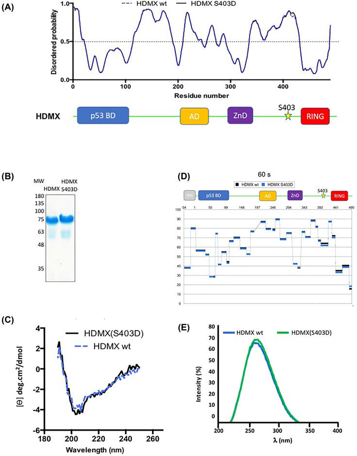
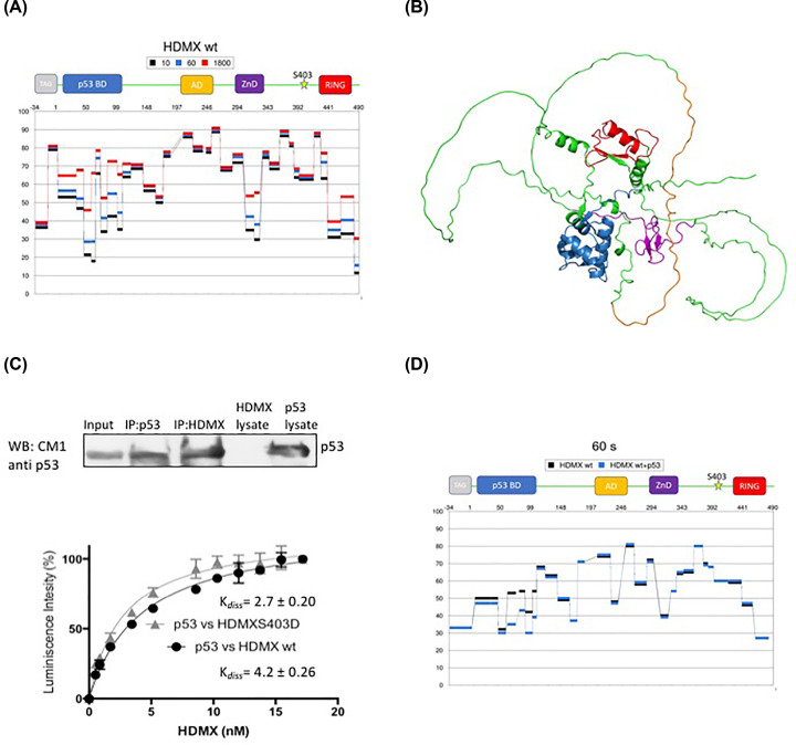
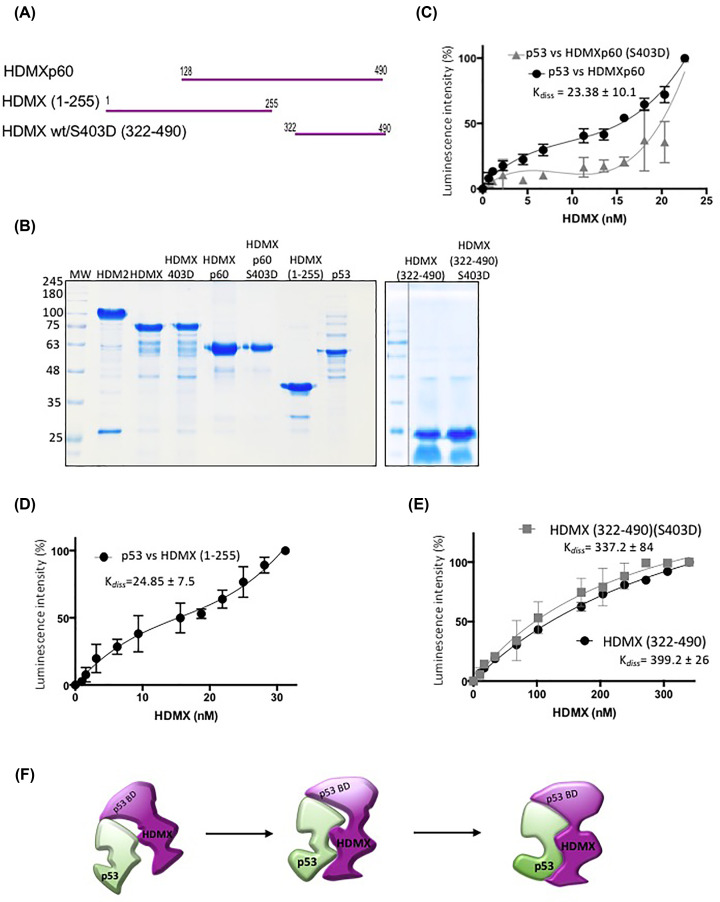
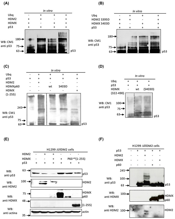
HDMX and its homologue HDM2 are two essential proteins for the cell; after genotoxic stress, both are phosphorylated near to their RING domain, specifically at serine 403 and 395, respectively. Once phosphorylated, both can bind the p53 mRNA and enhance its translation; however, both recognize p53 protein and provoke its degradation under normal conditions. HDM2 has been well-recognized as an E3 ubiquitin ligase, whereas it has been reported that even with the high similarity between the RING domains of the two homologs, HDMX does not have the E3 ligase activity. Despite this, HDMX is needed for the proper p53 poly-ubiquitination. Phosphorylation at serine 395 changes the conformation of HDM2, helping to explain the switch in its activity, but no information on HDMX has been published. Here, we study the conformation of HDMX and its phospho-mimetic mutant S403D, investigate its E3 ligase activity and dissect its binding with p53. We show that phospho-mutation does not change the conformation of the protein, but HDMX is indeed an E3 ubiquitin ligase in vitro; however, in vivo, no activity was found. We speculated that HDMX is regulated by induced fit, being able to switch activity accordingly to the specific partner as p53 protein, p53 mRNA or HDM2. Our results aim to contribute to the elucidation of the contribution of the HDMX to p53 regulation.

Keywords: HDM2; HDMX; Induced fit; MDM2; MDMX; cancer; p53; ubiquitination.


Conflict of interest statement

The authors declare that there are no competing interests associated with the manuscript.

Figures

Figure 1
Figure 1. HDMX does not show conformational rearrangement by phosphomimetic mutant HDMX(S403D)
(A) The predicted intrinsically disordered regions of HDMX (dotted blue line) and HDMX (S403D) (black line) are shown in the upper panel [28]. Cartoon illustrating major domains of HDMX; p53 BD, the p53 binding domain (blue); AD, acidic domain (yellow); ZnD, zinc finger domain (violet); RING, really interesting new gene domain (red) the star represents the place of the phosphomimetic mutation S403D. (B) Recombinant purified HDMX and HDMX(S403D). MW, molecular weights in kDa. (C) Far-UV CD spectra of the HDMX and phosphomimetic mutant HDMX(S403D). (D) HDMX and HDMX (S403D) mutant difference in deuteration, the data are plotted as % of deuteration of the peptide as a function of the numbering of the amino acids -34–490 after 60 s of incubation in the deuterated buffer. The -34 to 1 is due to the 6XHIS tag. For other incubation times, see Supplementary Figure S1. (E) The intrinsic fluorescence emission spectra of HDMX and phosphomimetic mutant HDMX(S403D). The spectra shown were obtained after subtracting the blank (no enzyme) from the experimental values. One representative experiment of three is shown.
Figure 2
Figure 2. HDMX is a very flexible protein
(A) HDX-MS of HDMX at different deuteration times, the data are plotted as % of deuteration of the peptide as a function of the numbering of the amino acids -34–490 after 10 s (in black), 60 s (in blue) and 1800 s (in red) of incubation in the deuterated buffer. The data shown represent the averages and standard deviations (SD) from five independent experiments. (B) Molecular modeling of HDMX using the AlphaFold Protein Structure Database (PMID: 34265844, PMID: 34791371) [30,31]; the p53 BD is shown in blue, the zinc finger in violet and RING in red are structurally ordered whereas the acidic domain in orange is part of the intrinsically disordered, other IRG are shown in green. (C) In the upper panel; recombinant p53 and HDMX proteins were synthesized from E. coli by separate. Then, a mix of equivalent protein quantities was used for immunoprecipitation with anti-p53 and anti-HDMX. Note that anti-p53 does not react with HDMX lysate. Lower panel; ELISA using a fixed amount of recombinant purified p53 (10 ng/μl) and increasing amounts of HDMX or HDMX(S403D) (0–20 ng/μl). HDMX and HDMX(S403D) show similar affinity towards p53, with Kdiss values of 4.4 and 2.7, respectively. (D) HDX MS of HDMX (in black) and HDMX-p53 interaction (in blue). The data are plotted as % of deuteration of the peptide as a function of the numbering of the amino acids (-34 to 1 is due to the 6XHis Tag) 1–490 after 60 s of incubation in the deuterated buffer.
Figure 3
Figure 3. The different domains of HDMX bind p53 protein
(A) The different construct using to dissect the interaction with p53. (B) SDS-PAGE of the recombinant purified constructs of HDMX, p53 and HDM2; MW, molecular weights in kilodaltons. (C) ELISA using a fixed amount of recombinant purified p53 (10 ng/μl) and increasing amounts of HDMXp60 or HDMXp60(S403D) (0–20 ng/μl). (D) ELISA using a fixed amount of recombinant purified p53 (10 ng/μl) and increasing amounts of HDMX(1-255). (E) ELISA using a fixed amount of recombinant purified p53 (10 ng/μl) and increasing amounts of HDMX (322–490) or HDMX (322–490)(S403D) (0–20 ng/μl). (F) Model of interaction between HDMX and p53 showing the flexibility of HDMX and the induced fix interaction.
Figure 4
Figure 4. Cryptic HDMX ubiquitin ligase activity towards p53
(A) In vitro ubiquitination of recombinant p53 with HDM2 and/or HDMX. (B) In vitro ubiquitination of recombinant p53 with HDM2(S395D) and/or HDMX(S403D). (C) In vitro ubiquitination of recombinant p53 with HDM2 or HDMXp60 and the phosphomimetic mutant, and HDMX (1-255). (D) In vitro ubiquitination of recombinant p53 with HDMX (322-490) wt and the phosphomimetic mutant. (E) Levels of expression of p53 in H1299 ∆HDM2 cell line transfected with the HDM2, HDMX, HDMXp60 and HDMX (1-255). (F) In vivo ubiquitination in H1299 ∆HDM2 cell line of p53 with HDM2, HDMX or HDMXp60 using MG 132 to inhibits proteasome activity.

Similar articles

References

    1. Li C., Chen L. and Chen J. (2002) DNA damage induces MDMX nuclear translocation by p53-dependent and -independent mechanisms. Mol. Cell. Biol. 22, 7562–7571 10.1128/MCB.22.21.7562-7571.2002 - DOI - PMC - PubMed
    1. Chen L., Gilkes D.M., Pan Y., Lane W.S. and Chen J. (2005) ATM and Chk2-dependent phosphorylation of MDMX contribute to p53 activation after DNA damage. EMBO J. 24, 3411–3422 10.1038/sj.emboj.7600812 - DOI - PMC - PubMed
    1. Malbert-Colas L., Ponnuswamy A., Olivares-Illana V., Tournillon A.S., Naski N. and Fahraeus R. (2014) HDMX folds the nascent p53 mRNA following activation by the ATM kinase. Mol. Cell. 54, 500–511 10.1016/j.molcel.2014.02.035 - DOI - PubMed
    1. Fahraeus R. and Olivares-Illana V. (2014) MDM2’s social network. Oncogene 33, 4365–4376 10.1038/onc.2013.410 - DOI - PubMed
    1. Gajjar M., Candeias M.M., Malbert-Colas L., Mazars A., Fujita J., Olivares-Illana V.et al. . (2012) The p53 mRNA-Mdm2 interaction controls Mdm2 nuclear trafficking and is required for p53 activation following DNA damage. Cancer Cell. 21, 25–35 10.1016/j.ccr.2011.11.016 - DOI - PubMed

Publication types

MeSH terms