. 2022 Jun 29;10(3):e0088422.
doi: 10.1128/spectrum.00884-22. Epub 2022 Jun 8.

Crizotinib Shows Antibacterial Activity against Gram-Positive Bacteria by Reducing ATP Production and Targeting the CTP Synthase PyrG

Affiliations

Crizotinib Shows Antibacterial Activity against Gram-Positive Bacteria by Reducing ATP Production and Targeting the CTP Synthase PyrG

Yun-Dan Zheng et al. Microbiol Spectr. .

Abstract

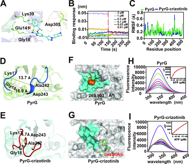
Infections caused by drug-resistant bacteria are a serious threat to public health worldwide, and the discovery of novel antibacterial compounds is urgently needed. Here, we screened an FDA-approved small-molecule library and found that crizotinib possesses good antimicrobial efficacy against Gram-positive bacteria. Crizotinib was found to increase the survival rate of mice infected with bacteria and decrease pulmonary inflammation activity in an animal model. Furthermore, it showed synergy with clindamycin and gentamicin. Importantly, the Gram-positive bacteria showed a low tendency to develop resistance to crizotinib. Mechanistically, quantitative proteomics and biochemical validation experiments indicated that crizotinib exerted its antibacterial effects by reducing ATP production and pyrimidine metabolism. A drug affinity responsive target stability study suggested crizotinib targets the CTP synthase PyrG, which subsequently disturbs pyrimidine metabolism and eventually reduces DNA synthesis. Subsequent molecular dynamics analysis showed that crizotinib binding occurs in close proximity to the ATP binding pocket of PyrG and causes loss of function of this CTP synthase. Crizotinib is a promising antimicrobial agent and provides a novel choice for the development of treatment for Gram-positive infections. IMPORTANCE Infections caused by drug-resistant bacteria are a serious problem worldwide. Therefore, there is an urgent need to find novel drugs with good antibacterial activity against multidrug-resistant bacteria. In this study, we found that a repurposed drug, crizotinib, exhibits excellent antibacterial activity against drug-resistant bacteria both in vivo and in vitro via suppressing ATP production and pyrimidine metabolism. Crizotinib was found to disturb pyrimidine metabolism by targeting the CTP synthase PyrG, thus reducing DNA synthesis. This unique mechanism of action may explain the decreased development of resistance by Staphylococcus aureus to crizotinib. This study provides a potential option for the treatment of drug-resistant bacterial infections in the future.

Keywords: Gram-positive bacteria; crizotinib; drug repurposing; quantitative proteomics.


Conflict of interest statement

The authors declare no conflict of interest.

Figures

FIG 1
FIG 1
Screening of an FDA-approved small-molecule library led to the identification of crizotinib as a prospective antibacterial agent. (A) Flow chart of screening antibacterial compound in the FDA-approved drug library; (B) MIC of crizotinib against Gram-positive and Gram-negative bacteria. The MICs of E. coli BW25113 and P. aeruginosa PA101 were larger than the maximal drug concentration (120 μg/mL) tested. (C) MIC of crizotinib against the monoresistant strains; (D) MIC of crizotinib against clinically isolated multiple-resistant strains. Brown represents clinically isolated multiple-resistant S. aureus, black represents clinically isolated multiple-resistant E. faecalis, yellow represents clinically isolated multiple-resistant S. haemolyticus 0078, and gray represents clinically isolated multiple-resistant E. coli. The MIC of clinically isolated multiple-resistant E. coli was larger than the maximal drug concentration (120 μg/mL) tested. (E) Growth curve of S. aureus cultured in TSB medium with 0× the MIC, 0.5× the MIC, and 1× the MIC of crizotinib; (F, G) SEM images of S. aureus exposed to DMSO (control) or 1× the MIC of crizotinib; (H) development of resistance of S. aureus Newman to crizotinib and Amp; (I) synergistic antibacterial efficacy of crizotinib combined with either Gent or Cli for S. aureus.
FIG 2
FIG 2
Therapeutic efficacy of crizotinib for the mice with MRSA-166138-induced pneumonia. (A) Survival rate of mice (n = 6); (B) CFU counts in the lung tissues at 48 h postinfection; (C) hematoxylin and eosin (HE) staining of lung collected from control group and crizotinib- and vehicle-treated groups. The arrow indicates alveoli with inflammatory cells. Scale bar, 100 μm; (D) pulmonary inflammation recorded through CT scanning. The arrow indicates pulmonary shadow; (E) HE staining of liver and kidney collected from crizotinib- and vehicle-treated mice; (F) ALT level in serum of the crizotonib- and vehicle-treated mice. *, P < 0.05; **, P < 0.01; ****, P < 0.0001.
FIG 3
FIG 3
Statistical analysis of proteomic changes in S. aureus Newman induced by crizotinib. (A) PLGEM fitting based on the abundance of crizotinib-regulated proteins; (B) volcano plots of total protein in the 1-h group and 2-h group; (C) numbers of upregulated and downregulated DEPs in the 1-h group and 2-h group; (D) diagram of numbers of DEPs in the 1-h group and 2-h group; (E) KEGG enrichment analysis of DEPs.
FIG 4
FIG 4
Crizotinib interferes with pyrimidine metabolism to decrease DNA synthesis. (A) Diagram showing the DEPs in citrate cycle; (B, C) acetyl-CoA and ATP levels of control S. aureus and S. aureus treated with 0.5× the MIC of crizotinib; (D) diagram showing the DEPs in pyrimidine metabolism; (E) RT-qPCR analysis of selected genes in S. aureus with crizotinib treatments for 1 h and 2 h versus that with the corresponding control group; (F) fluorescence histogram of DAPI in S. aureus with (deep purple) or without (light brown) treatment with 0.5× the MIC of crizotinib; (G) MFI of DAPI in S. aureus with or without treatment with 0.5× the MIC of crizotinib. *, P < 0.05; **, P < 0.01.
FIG 5
FIG 5
Crizotinib directly targets PyrG. (A) Schematic diagram of DARTS technology; (B) pronase digestion of the total proteins of S. aureus Newman incubated with crizotinib or DMSO; (C to E) DARTS, thermal stability, and BLI assays showing the binding between crizotinib and PyrG; (F) species evolutionary tree; (G) growth inhibition by crizotinib of the D39 WT and D39 Δpyrg strains in 2, 4, and 6 h; (H) growth inhibition by crizotinib of the D39 ΔpyrG::pyrG and D39 ΔpyrG::pyrGG18A K39A E141A D305A strains in 4 and 6 h; (I) OD600 determined after supplementation with a series of CTP concentrations of S. aureus Newman treated with 0.5× the MIC of crizotinib. Error bars indicate SD. *, P < 0.05; **, P < 0.01; ***, P < 0.001.
FIG 6
FIG 6
Crizotinib binding induced a conformational change of PyrG. (A) Schematic representation of the interactions between PyrG and crizotinib analyzed by UCSF Dock. Asp305, Gly18, Glu141, and Lys39 are located in the binding site. (B) BLI assay of the binding between crizotinib and PyrGG18A K39A E141A D305A; (C) RMSF values of all residues of PyrG-crizotinib and PyrG proteins calculated over the 100-ns trajectory; (D, E) distances between ATP-binding residues in PyrG (navy) and PyrG-crizotinib complex (red) measured by PyMOL; (F, G) closing of the ATP binding pocket in PyrG induced by crizotinib binding. The residues of the ATP binding pocket in PyrG and PyrG-crizotinib complex are colored in blue. The structures were visualized at 90 ns during molecular dynamics simulation; (H, I) fluorescence titration showing the interaction between ATP and PyrG-crizotinib complex or PyrG.
FIG 7
FIG 7
Diagram showing that crizotinib inhibits S. aureus by reducing ATP production and targeting PyrG in pyrimidine metabolism and then decreasing DNA synthesis.

References

    1. Cassini A, Högberg LD, Plachouras D, Quattrocchi A, Hoxha A, Simonsen GS, Colomb-Cotinat M, Kretzschmar ME, Devleesschauwer B, Cecchini M, Ouakrim DA, Oliveira TC, Struelens MJ, Suetens C, Monnet DL, Strauss R, Mertens K, Struyf T, Catry B, Latour K, Ivanov IN, Dobreva EG, Tambic Andraševic A, Soprek S, Budimir A, Paphitou N, Žemlicková H, Schytte Olsen S, Wolff Sönksen U, Märtin P, Ivanova M, Lyytikäinen O, Jalava J, Coignard B, Eckmanns T, Abu Sin M, Haller S, Daikos GL, Gikas A, Tsiodras S, Kontopidou F, Tóth Á, Hajdu Á, Guólaugsson Ó, Kristinsson KG, Murchan S, Burns K, Pezzotti P, Gagliotti C, Dumpis U, et al. . 2019. Attributable deaths and disability-adjusted life-years caused by infections with antibiotic-resistant bacteria in the EU and the European Economic Area in 2015: a population-level modelling analysis. Lancet Infect Dis 19:56–66. doi:10.1016/S1473-3099(18)30605-4. - DOI - PMC - PubMed
    1. Chambers HF, Deleo FR. 2009. Waves of resistance: Staphylococcus aureus in the antibiotic era. Nat Rev Microbiol 7:629–641. doi:10.1038/nrmicro2200. - DOI - PMC - PubMed
    1. Tacconelli E, Carrara E, Savoldi A, Harbarth S, Mendelson M, Monnet DL, Pulcini C, Kahlmeter G, Kluytmans J, Carmeli Y, Ouellette M, Outterson K, Patel J, Cavaleri M, Cox EM, Houchens CR, Grayson ML, Hansen P, Singh N, Theuretzbacher U, Magrini N, Aboderin AO, Al-Abri SS, Awang Jalil N, Benzonana N, Bhattacharya S, Brink AJ, Burkert FR, Cars O, Cornaglia G, Dyar OJ, Friedrich AW, Gales AC, Gandra S, Giske CG, Goff DA, Goossens H, Gottlieb T, Guzman Blanco M, Hryniewicz W, Kattula D, Jinks T, Kanj SS, Kerr L, Kieny M-P, Kim YS, Kozlov RS, Labarca J, Laxminarayan R, Leder K, WHO Pathogens Priority List Working Group, et al. . 2018. Discovery, research, and development of new antibiotics: the WHO priority list of antibiotic-resistant bacteria and tuberculosis. Lancet Infect Dis 18:318–327. doi:10.1016/S1473-3099(17)30753-3. - DOI - PubMed
    1. Lowy FD. 1998. Staphylococcus aureus infections. N Engl J Med 339:520–532. doi:10.1056/NEJM199808203390806. - DOI - PubMed
    1. Tong SYC, Davis JS, Eichenberger E, Holland TL, Fowler VG, Jr.. 2015. Staphylococcus aureus infections: epidemiology, pathophysiology, clinical manifestations, and management. Clin Microbiol Rev 28:603–661. doi:10.1128/CMR.00134-14. - DOI - PMC - PubMed

Publication types