. 2022 Jun 6;27(1):e22spe1.
doi: 10.1590/2177-6709.27.1.e22spe1. eCollection 2022.

Agenesis of maxillary lateral incisors: diagnosis and treatment options

Affiliations

Agenesis of maxillary lateral incisors: diagnosis and treatment options

Daniela Kimaid Schroeder et al. Dental Press J Orthod. .

Abstract

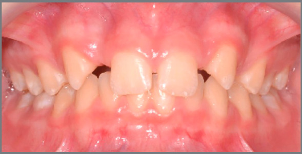
Introduction: There are different possibilities of orthodontic planning for cases with congenital absence of maxillary lateral incisors. This subject divides the opinion of orthodontists and oral rehabilitation clinicians, due to the advantages and disadvantages of each treatment option, which may involve opening spaces for future implants and/or prosthetic restorations, or closing the spaces by positioning the maxillary canines in the place of lateral incisors. The correct diagnosis and careful evaluation of each patient allow to determine the best therapeutic approach. This paper discusses the main topics to be considered when planning these cases.

Objectives: To evaluate the main aspects related to orthodontic treatment planning in cases of congenital absence of maxillary lateral incisors, to aid the decision-making, with clinical and scientific basis.

Introdução:: Existem diferentes possibilidades de planejamento ortodôntico para os casos que apresentam ausência congênita de incisivos laterais superiores. Esse é um assunto que divide a opinião de ortodontistas e reabilitadores orais, devido às vantagens e desvantagens de cada uma das opções de tratamento, as quais podem envolver a abertura de espaços para futuros implantes e/ou restaurações protéticas ou o fechamento dos espaços, com posicionamento dos caninos superiores no lugar dos incisivos laterais. O correto diagnóstico e uma criteriosa avaliação de cada paciente permitem determinar a melhor abordagem terapêutica. Nesse artigo, serão discutidos os principais tópicos a serem considerados no planejamento desses casos.

Objetivos:: Avaliar os principais aspectos relacionados ao planejamento do tratamento ortodôntico nos casos de ausência congênita de incisivos laterais superiores, de maneira a auxiliar nas tomadas de decisão, com embasamento clínico e científico.


Conflict of interest statement

The authors report no commercial, proprietary or financial interest in the products or companies described in this article.

Figures

Figure 1:
Figure 1:. 11-year-old female patient with esthetic complaint due to absence of maxillary lateral incisors.
Figure 2:
Figure 2:. Smile at 13 years of age. Case treated with space closure, with maxillary canines replacing the missing maxillary lateral incisors.
Figure 3:
Figure 3:. Adult patient, with absence of maxillary lateral incisors, maxillary anterior spaces and mandibular crowding. Class II canine relationship, mild overbite and overjet.
Figure 4:
Figure 4:. Case treated with space opening for replacement of maxillary lateral incisors with dental implants.
Figure 5:
Figure 5:. Hook made with stainless steel wire, fitted to the distal part of the tube of tooth #17, connected to the mini-implant between the roots of teeth #14 and #15 using a chain elastic, aiming at mesialization of the posterior segment, closing the spaces by anchorage loss.
Figure 6:
Figure 6:. A) Image of adult patient with low smile, after orthodontic space opening for implant placement in the regions of maxillary lateral incisors. B) The presence of bone dehiscence on the buccal walls of teeth #12 and #22 could compromise the smile esthetics, if there was any degree of gingival exposure.
Figure 7:
Figure 7:. A, B) Lateral intraoral photographs of initial occlusion, with molars and canines in Class II relationship, congenital absence of maxillary lateral incisors, generalized spaces in the maxillary arch. C, D) Post-orthodontic treatment photographs showing closure of spaces, with mesialization of maxillary posterior teeth. Teeth #13 and #23 replaced the missing teeth #12 and #22, and the molar relationship settled into a full Class II relationship.
Figure 8:
Figure 8:. A, B) Posterior occlusion on the right and left sides, periodontal health in the premolar region with 7-year follow-up. C) Follow-up panoramic radiograph.
Figure 9:
Figure 9:. A, B) Occlusion after completion of orthodontic treatment, performed by replacing the congenitally absent maxillary lateral incisors with maxillary canines. C) Panoramic radiograph at treatment completion.
Figure 10:
Figure 10:. Photographs taken five years after treatment completion, showing a stable occlusion, besides dental and periodontal health.
Figure 11:
Figure 11:. A) Frontal view of a case of congenital absence of teeth #12 and #22 orthodontically treated by space closure. B) Ten years after treatment completion, the patient started the tooth whitening process. C) Right lateral view of the final occlusion. D) Image ten years after treatment. E) Left lateral view of the final occlusion. F) The same image after ten years of treatment, evidencing on the photographs taken in the post-retention phase that the periodontium and anatomy of premolars involved with the group lateral disocclusion guidance remained healthy.
Figure 12:
Figure 12:. Young patient, aged 9 years at completion of mixed dentition, with molar and canine relationship in key occlusion, posterior crossbite on the left side, absence of tooth #12 and microdontia associated with a peg-shaped crown of tooth #22. The maxillary left canine had a radiographic appearance of possible impaction.
Figure 13:
Figure 13:. Images of occlusion after orthodontic treatment performed by closing the maxillary spaces. Expansion of maxillary arch and extraction of the peg-shaped lateral incisor were performed. Anatomical re-shaping was performed on the permanent maxillary canines, aiming at improving the esthetics and function.
Figure 14:
Figure 14:. Images of the case, 8 years after completion of orthodontic treatment. Esthetic completion of the case was performed by restorative dentistry, by re-shaping of maxillary incisors, canines and first premolars.
Figure 15:
Figure 15:. Smile images: A) Initial; B) After completion of orthodontic treatment, in which it is possible to observe the change in color and anatomy of canines; C) After esthetic restorative procedures.
Figure 16:
Figure 16:. Canines with palatal root torque and slightly mesially rotated maxillary first premolars.
Figure 17:
Figure 17:. Retainer bonded to central incisors and canines, to increase treatment stability and prevent reopening of small spaces.

References

    1. Pinho T, Maciel P, Pollmann C. Developmental disturbances associated with agenesis of the permanent maxillary lateral incisor. Br Dent J. 2009;207(12):E25. - PubMed
    1. Pinho T, Tavares P, Maciel P, Pollmann C. Developmental absence of maxillary lateral incisors in the Portuguese population. Eur J Orthod. 2005;27(5):443–449. - PubMed
    1. Turpin DL. Treatment of missing lateral incisors. Am J Orthod Dentofacial Orthop. 2004;125(2):129–129. - PubMed
    1. Olsen TM, Kokich VG., Sr Postorthodontic root approximation after opening space for maxillary lateral incisor implants. Am J Orthod Dentofacial Orthop. 2010;137(2):158–159. - PubMed
    1. Zachrisson BU, Rosa M, Toreskog S. Congenitally missing maxillary lateral incisors: canine substitution. Point. Am J Orthod Dentofacial Orthop. 2011;139(4):434,436,438. - PubMed