Review
. 2022 Oct;49(12):4133-4144.
doi: 10.1007/s00259-022-05849-y. Epub 2022 Jun 8.

CXCR4-targeted theranostics in oncology

Affiliations
Review

CXCR4-targeted theranostics in oncology

Andreas K Buck et al. Eur J Nucl Med Mol Imaging. 2022 Oct.

Abstract

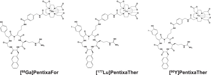
A growing body of literature reports on the upregulation of C-X-C motif chemokine receptor 4 (CXCR4) in a variety of cancer entities, rendering this receptor as suitable target for molecular imaging and endoradiotherapy in a theranostic setting. For instance, the CXCR4-targeting positron emission tomography (PET) agent [68 Ga]PentixaFor has been proven useful for a comprehensive assessment of the current status quo of solid tumors, including adrenocortical carcinoma or small-cell lung cancer. In addition, [68 Ga]PentixaFor has also provided an excellent readout for hematological malignancies, such as multiple myeloma, marginal zone lymphoma, or mantle cell lymphoma. PET-based quantification of the CXCR4 capacities in vivo allows for selecting candidates that would be suitable for treatment using the theranostic equivalent [177Lu]/[90Y]PentixaTher. This CXCR4-directed theranostic concept has been used as a conditioning regimen prior to hematopoietic stem cell transplantation and to achieve sufficient anti-lymphoma/-tumor activity in particular for malignant tissues that are highly sensitive to radiation, such as the hematological system. Increasing the safety margin, pretherapeutic dosimetry is routinely performed to determine the optimal activity to enhance therapeutic efficacy and to reduce off-target adverse events. The present review will provide an overview of current applications for CXCR4-directed molecular imaging and will introduce the CXCR4-targeted theranostic concept for advanced hematological malignancies.

Keywords: Adrenocortical carcinoma; C-X-C motif chemokine receptor 4; CXCR4; Endoradiotherapy; Multiple myeloma; Theranostics; [177Lu]PentixaTher; [68Ga]PentixaFor; [90Y]PentixaTher.


Conflict of interest statement

The authors declare no competing interests.

Figures

Fig. 1
Fig. 1
Chemical structures of [68Ga]PentixaFor, [90Y], and [.177Lu]PentixaTher
Fig. 2
Fig. 2
Patient after resection of adrenocortical carcinoma imaged with [68 Ga]PentixaFor. The right-sided primary was resected earlier. Maximum intensity projection in A revealed multiple sites of disease after administration of [68Ga]PentixaFor. Transaxial CT (B), PET (C), and PET/CT (D) demonstrated intense uptake in a retroperitoneal lesion. Further CXCR4 positive sites of disease included liver lesions (E and G). Additional discernible uptake in the left adrenal gland (F)
Fig. 3
Fig. 3
Patient with marginal zone lymphoma after injection of [68 Ga]PentixaFor. Multiple disease sites are visualized on maximum intensity projection in A. Transaxial CT (B), PET (C), and PET/CT (D) revealed intense lymph node manifestations in the thorax. PET/CT also showed radiotracer accumulation in the cervical (E), abdominal (F), and in the inguinal region (G). Modified from Duell et al., Journal of Nuclear Medicine, October 2021, 62 (10) 1415–1421 [11]. © by the Society of Nuclear Medicine and Molecular Imaging, Inc
Fig. 4
Fig. 4
Bar chart showing A average SUVmax and B target-to-background ratio (TBR). For A, black dotted lines indicate SUVmax cutoffs of 6 and 12, and for B, those lines show TBR cutoffs of 4 and 8, respectively. BP blood pool (red dotted line), AML acute myeloid leukemia, CCC cholangiocarcinoma, NSCLC non-small-cell lung carcinoma, NEN neuroendocrine neoplasm, DSRCT Desmoplastic Small Round Cell Tumor, ALL acute lymphoblastoid leukemia, CLL chronic lymphocytic leukemia, MZL marginal zone lymphoma, SCLC small-cell lung carcinoma, MM multiple myeloma. Adrenocortical adenoma: aldosteron-producing adrenocortical adenoma. Modified from Buck et al., Journal of Nuclear Medicine, 2022 Mar 3; jnumed.121.263693 [43]. © by the Society of Nuclear Medicine and Molecular Imaging, Inc
Fig. 5
Fig. 5
Example of activity time functions in a patient with multiple myeloma. Activity retention measurements as well as fit functions are shown for the whole body (black), per liter of whole blood (grey), red bone marrow (red), liver (green), kidneys (purple), and spleen (blue). Modified from Hänscheid et al., Journal of Nuclear Medicine 2021 Aug 19; jnumed.121.262295, https://doi.org/10.2967/jnumed.121.262295 [46]. © by the Society of Nuclear Medicine and Molecular Imaging, Inc
Fig. 6
Fig. 6
Partial response in a patient affected with diffuse large B cell lymphoma treated with CXCR4-targeted endoradiotherapy and additional radioimmunotherapy. Pretherapeutic [68Ga]PentixaFor (left) and posttherapeutic [18F]FDG PET/CT (right) after tandem treatment using [90Y]PentixaTher and [90Y]Ibritumomab-Tiuxetan (Zevalin). Posttherapy scans demonstrated reduction of lesions in the kidneys, adrenals (arrows), lung, and nodal disease manifestations. Note that diffuse radiotracer accumulation in the lung on [18F]FDG maximum intensity projection on the right was due to pneumonia. Modified from Lapa et al., Journal of Nuclear Medicine Jan 2019, 60 (1) 60–64 [15], © by the Society of Nuclear Medicine and Molecular Imaging, Inc
Fig. 7
Fig. 7
A Reduction of blood values relative to baseline after CXCR4-directed endoradiotherapy. B Time interval between CXCR4-directed endoradiotherapy and start of conventional conditioning therapy. Desired cytopenia was achieved for both [90Y]PentixaTher and [177Lu]PentixaTher (A). For [90Y]PentixaTher, however, the time interval until start of conditioning therapy was significantly shorter, which can be explained by the longer half-life of 2.7 days (177Lu, 6.7 days) (B). Modified from Maurer et al., 2019 Oct; 60(10): 1399–1405 [23], © by the Society of Nuclear Medicine and Molecular Imaging, Inc

References

    1. Chatterjee S, Behnam Azad B, Nimmagadda S. The intricate role of CXCR4 in cancer. Adv Cancer Res. 2014;124:31–82. doi: 10.1016/B978-0-12-411638-2.00002-1. - DOI - PMC - PubMed
    1. Demmer O, Gourni E, Schumacher U, Kessler H, Wester HJ. PET imaging of CXCR4 receptors in cancer by a new optimized ligand. ChemMedChem. 2011;6:1789–1791. doi: 10.1002/cmdc.201100320. - DOI - PMC - PubMed
    1. Herrmann K, Lapa C, Wester HJ, Schottelius M, Schiepers C, Eberlein U, et al. Biodistribution and radiation dosimetry for the chemokine receptor CXCR4-targeting probe 68Ga-pentixafor. J Nucl Med. 2015;56:410–416. doi: 10.2967/jnumed.114.151647. - DOI - PubMed
    1. Hartimath SV, Domanska UM, Walenkamp AM, Rudi AJOD, de Vries EF. [(9)(9)mTc]O(2)-AMD3100 as a SPECT tracer for CXCR4 receptor imaging. Nucl Med Biol. 2013;40:507–517. doi: 10.1016/j.nucmedbio.2013.02.003. - DOI - PubMed
    1. Nimmagadda S, Pullambhatla M, Pomper MG. Immunoimaging of CXCR4 expression in brain tumor xenografts using SPECT/CT. J Nucl Med. 2009;50:1124–1130. doi: 10.2967/jnumed.108.061325. - DOI - PMC - PubMed