

Correlation of somatostatin receptor PET/CT imaging features and immunohistochemistry in neuroendocrine tumors of the lung: a retrospective observational study
- PMID: 35674739
- PMCID: PMC9525360
- DOI: 10.1007/s00259-022-05848-z
Correlation of somatostatin receptor PET/CT imaging features and immunohistochemistry in neuroendocrine tumors of the lung: a retrospective observational study
Erratum in
-
Correction to: Correlation of somatostatin receptor PET/CT imaging features and immunohistochemistry in neuroendocrine tumors of the lung: a retrospective observational study.Eur J Nucl Med Mol Imaging. 2022 Oct;49(12):4289. doi: 10.1007/s00259-022-05877-8. Eur J Nucl Med Mol Imaging. 2022. PMID: 35699752 Free PMC article. No abstract available.
Abstract
Purpose: To correlate somatostatin receptor (SSTR) and proliferative activity profile (SSTR2, SSTR5, Ki-67) at immunohistochemistry (IHC) with SSTR-PET/CT imaging features in a retrospective series of lung neuroendocrine tumors (NET). Proliferative activity by Ki-67 and 18F-FDG-PET/CT parameters (when available) were also correlated.
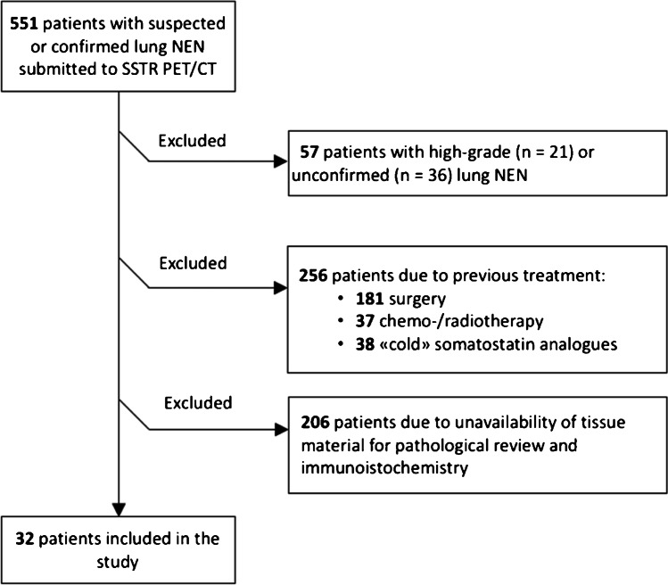
Methods: Among 551 patients who underwent SSTR-PET/CT with 68Ga-DOTA-somatostatin analogs (SSA) between July 2011 and March 2020 for lung neuroendocrine neoplasms, 32 patients with a confirmed diagnosis of NET were included. For 14 of them, 18F-FDG-PET/CT was available. PET/CT images were reviewed by qualitative and semi-quantitative analyses. Immunohistochemistry for SSTR2, SSTR5, and Ki-67 was assessed. Inferential analysis was performed including kappa statistics and Spearman's rank correlation test.
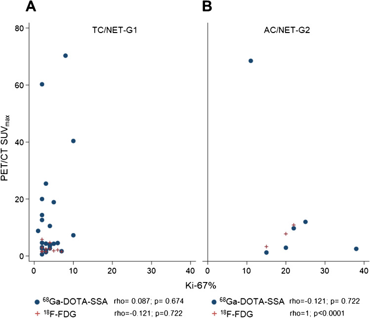
Results: Definitive diagnosis consisted of 26 typical carcinoids-G1 and six atypical carcinoids-G2. Positive SSTR2-IHC was found in 62.5% of samples while SSTR5-IHC positivity was 19.4%. A correlation between SSTR2-IHC and SSTR-PET/CT was found in 24/32 cases (75.0%, p = 0.003): 20 were concordantly positive, 4 concordantly negative. For positive IHC, 100% concordance with SSTR-PET/CT (both positive) was observed, while for negative IHC concordance (both negative) was 33.3%. In 8 cases, IHC was negative while SSTR-PET/CT was positive, even though with low-grade uptake in all but one. A significant correlation between SUVmax values at SSTR-PET/CT and the SSTR2-IHC scores was found, with low SUVmax values corresponding to negative IHC and higher SUVmax values to positive IHC (p = 0.002).
Conclusion: This retrospective study showed an overall good agreement between SSTR2-IHC and tumor uptake at SSTR-PET/CT in lung NETs. SSTR-PET/CT SUVmax values can be used as a parameter of SSTR2 density. Within the limits imposed by the relatively small cohort, our data suggest that SSTR2-IHC may surrogate SSTR-PET/CT in selected lung NET patients for clinical decision making when SSTR-PET/CT is not available.
Keywords: Immunohistochemistry; Ki-67; Lung NET; PET/CT; Somatostatin receptor subtypes; [68Ga]-DOTA-somatostatin analogs.
© 2022. The Author(s).
Conflict of interest statement
Financial interests: Filippo Lococo and Stefano Margaritora received speaker and consultant honoraria from Astrazeneca. Guido Rindi received speaker and consultant honoraria from Company AAA and Bracco.
Vittoria Rufini, Margherita Lorusso, Frediano Inzani, Tina Pasciuto, Elizabeth KA Triumbari, Lucia Rosalba Grillo, and Edoardo Pescarmona declare no financial interests.
Figures


References
-
- Rindi G, Klimstra DS, Abedi-Ardekani B, Asa SL, Bosman FT, Brambilla E, et al. A common classification framework for neuroendocrine neoplasms: an International Agency for Research on Cancer (IARC) and World Health Organization (WHO) expert consensus proposal. Mod Pathol. 2018;31:1770–1786. doi: 10.1038/s41379-018-0110-y. - DOI - PMC - PubMed
-
- Travis WD, Beasley MB, Cree IA et al. Chapter 1.4.1: Lung neuroendocrine neoplasms: Introduction. In: WHO Classification of Tumours Editorial Board. Thoracic tumours. Lyon (France): International Agency for Research on Cancer; 2021. (WHO classification of tumours series, 5th ed.; vol. 5, pp. 127–129). https://publications.iarc.fr/595.
-
- Papotti M, Brambilla E, Dingemans AC et al. Chapter 1.4.3.1: Carcinoid/Neuroendocrine tumour of the lung. In: WHO Classification of Tumours Editorial Board. Thoracic tumours. Lyon (France): International Agency for Research on Cancer; 2021. (WHO classification of tumours series, 5th ed.; vol.5, pp. 133–8). https://publications.iarc.fr/595.
-
- Papotti M, Croce S, Bellò M, Bongiovanni M, Allìa E, Schindler M, et al. Expression of somatostatin receptor types 2, 3 and 5 in biopsies and surgical specimens of human lung tumours. Correlation with preoperative octreotide scintigraphy. Virchows Arch. 2001;439:787–97. doi: 10.1007/s004280100494. - DOI - PubMed
Publication types
MeSH terms
Substances
LinkOut - more resources
Full Text Sources
Medical
Research Materials
