. 2022 Jun 14;119(24):e2114309119.
doi: 10.1073/pnas.2114309119. Epub 2022 Jun 8.

Mosquito saliva enhances virus infection through sialokinin-dependent vascular leakage

Affiliations

Mosquito saliva enhances virus infection through sialokinin-dependent vascular leakage

Daniella A Lefteri et al. Proc Natl Acad Sci U S A. .

Abstract

Viruses transmitted by Aedes mosquitoes are an increasingly important global cause of disease. Defining common determinants of host susceptibility to this large group of heterogenous pathogens is key for informing the rational design of panviral medicines. Infection of the vertebrate host with these viruses is enhanced by mosquito saliva, a complex mixture of salivary-gland-derived factors and microbiota. We show that the enhancement of infection by saliva was dependent on vascular function and was independent of most antisaliva immune responses, including salivary microbiota. Instead, the Aedes gene product sialokinin mediated the enhancement of virus infection through a rapid reduction in endothelial barrier integrity. Sialokinin is unique within the insect world as having a vertebrate-like tachykinin sequence and is absent from Anopheles mosquitoes, which are incompetent for most arthropod-borne viruses, whose saliva was not proviral and did not induce similar vascular permeability. Therapeutic strategies targeting sialokinin have the potential to limit disease severity following infection with Aedes-mosquito-borne viruses.

Keywords: arbovirus; endothelium; inflammation; mosquitoes.


Conflict of interest statement

The authors declare no competing interest.

Figures

Fig. 1.
Fig. 1.
Mosquito saliva is sufficient to enhance virus infection and worsen clinical outcome. (AF) Mice were inoculated with either 104 PFU of SFV4 or 103 PFU of ZIKV into skin of left foot (upper side), alone or following Ae. aegypti mosquito biting, or coinjected with Aedes saliva normalized for total protein content (mosquito salivated 0.3714 μg of protein on average). Viral RNA and host 18S were quantified by qPCR and viral titers of serum by plaque assays at 24 hpi. (A) Mice were injected with SFV alone or alongside saliva from 1, 5, or 25 mosquitoes (n = 6). (B) Mice were injected with ZIKV with or without saliva from five mosquitoes (1.86 μg of protein, n = 6). (C) Mice were infected with SFV following 1 or 10 repeated tissue piercings with a hyperfine needle. (n = 6). (D) Survival of mice infected with 104 PFU of SFV4 (n = 10). (E and F) Mice were inoculated with SFV alone in resting skin, or into mosquito-bitten skin (5 bites), or into resting-skin mosquito saliva (1.86 μg of saliva protein, n = 8). *P < 0.05, **P < 0.01, ***P < 0.001, ns = not significant.
Fig. 2.
Fig. 2.
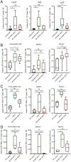
A heat-sensitive salivary factor from female mosquitoes enhances virus infection independent of bacterial microbiota. (A) Mouse skin was injected with 1.86 μg of saliva from control and antibiotic (Abx)-treated mosquitoes, and host expression of cxcl2, il1b, and ccl2 transcripts was assessed at 6 h (n = 6). (BD) Mouse skin was inoculated with 104 PFU of SFV4 alone or with Ae. aegypti saliva in the upper skin of the left foot. Viral RNA and host 18S were quantified from skin and spleen by qPCR and viral titers of serum by plaque assays at 24 hpi. (B) Female Ae. aegypti saliva from Abx-treated or untreated mosquitoes (n > 6). (C) Heat-treated (10 min at 95 °C) or untreated female Ae. aegypti saliva (n = 6). (D) Male or female Ae. aegypti saliva pooled from five mosquitoes combined, reared in the same cage (n = 6). *P < 0.05, **P < 0.01, ***P < 0.001, ns = not significant.
Fig. 3.
Fig. 3.
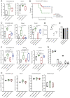
Anopheles mosquito saliva lacks the ability to enhance virus infection. (AC) Mouse skin was inoculated with either 105 PFU of ZIKV or 104 PFU of SFV4 alone or with 1.86 μg of saliva of either Ae. aegypti, An. gambiae, or An. stephensi. Virus RNA and host 18S and serum viral titers were quantified at 24 hpi. (B) Survival of mice infected with 4 × 10⁴ PFU of SFV4. (D) Macrophages were infected with luciferase-expressing SFV at an multiplicity of infection of 0.1 alone or with 0.66 μg of protein of Ae. aegypti, An. gambiae, or Ae. albpictus saliva. Luciferase activity of tissue culture media was assayed at 6 hpi (n = 6). (E) BHK cells were infected with 10-fold serial dilutions of SFV4 ranging between 25,000 PFU and 0.25 PFU alone or with Ae. aegypti or An. gambiae saliva and then immediately overlayed with Avicel. PFUs were then assessed at 48 hpi. Shown here are representative PFUs for wells in which plaques were quantifiable. (F) Mouse skin was inoculated with 104 PFU of SFV4 alone or alongside 1.86 μg of saliva of Ae. aegypti, An. gambiae, or both species. Virus RNA and host 18S were quantified from skin and spleen by qPCR and viral titers of serum by plaque assays at 24 hpi. (G and H) Mice were treated with 1.5 mg of anti-IFNAR antibodies (clone MAR1-5A3) and 24 h later were infected with 2 x10⁵ PFU ONNV s.c. in the skin (upper side of the left foot). (G) ONNV RNA quantities in tissues at 48 hpi were defined by qPCR to define tissue tropism. (H) Mouse skin was infected with ONNV alone or alongside 1.86 μg of saliva of either Ae. aegypti or An. gambiae. ONNV RNA and host 18S from tissues were quantified by qPCR, and serum viral titers were quantified via plaque assays at 48 hpi. *P < 0.05, **P < 0.01, ***P < 0.001, ****P < 0.0001, ns = not significant.
Fig. 4.
Fig. 4.
Immune sensing of Aedes saliva is not sufficient to enhance virus infection. (A and B) Mouse skin was injected with either saline control or 1.86 μg of saliva of either Ae. aegypti or An. gambiae. (A) Copy number of host transcripts in the skin was determined by qPCR at 6 h (n = 6). (B) Mice were inoculated with 104 PFU of SFV4 with either Ae. aegypti or An. gambiae saliva and transcripts quantified by qPCR (n = 8). (C) Mice treated with IFNAR-1 blocking antibody a day prior to inoculation with 10,000 PFU SFV4 into mouse skin (resting or following mosquito biting). Viral RNA and host 18S were quantified from skin and spleen by qPCR and viral titers of serum by plaque assays at 24 hpi. (n = 6). (D) Mosquito-bitten mouse skin was injected with saliva from five mosquitoes of either Ae. aegypti or An. gambiae. At 2 h, skin from the inoculation site was biopsied and digested to release cells, and numbers of myeloid cells (CD45+CD11b+), neutrophils (CD45+CD11b+Ly6G+Ly6Cint), and myelomonocytic cells (CD45+ CD11b+ Ly6G− Ly6C+ cells) were quantified (n = 6). (EG) BALB/c mouse skin was inoculated with 10,000 PFU of SFV alone or with saliva. Mice were either naive to saliva or primed to saliva by prior injections of mosquito saliva weekly for four consecutive weeks. (E) IL-13 transcript expression and cell numbers of draining popliteal lymph nodes at 2 hpi. IL-13 transcripts were quantified by qPCR (n = 6). (F) Virus RNA in skin was measured by qPCR and serum virus quantified by plaque assay, at 24 hpi (n = 6) for mice primed with either Aedes saliva or saline alone. (G) Mice were presensitized to either Aedes or Anopheles saliva for 4 wk prior to SFV infection coinjected with respective species saliva. Expression of the viral SFV gene was measured using qPCR in the skin and serum virus quantified by plaque assay at 24 hpi. *P < 0.05, **P < 0.01, ***P < 0.001, ****P < 0.0001, ns = not significant.
Fig. 5.
Fig. 5.
Increased vascular permeability induced by Aedes saliva enhances virus infection. (A and B) Mice administered i.p. with Evans blue were injected with 1.86 μg of mosquito saliva in the skin or exposed to up to three bites from Ae. aegypti or An. Gambiae. The extent of edema was assessed by quantification of Evan’s blue dye leakage into skin at 30 min and 3 h postsaliva/biting via colorimetric assay (n = 6). (C) Mouse skin was inoculated with 104 PFU of SFV4 alone or with either Ae. aegypti saliva or 10 μg of histamine dihydrochloride. SFV RNA and host 18S and serum viral titers were quantified at 24 hpi. (D) Mice were administered i.p with Evans blue and then given either control or antihistamines s.c. (0.5 mg cetirizine in 100 μL, 0.02 mg loratadine in 100 μL, and 0.1 mg of fexofenadine in 200 μL) 1 h prior to saliva injection. Mouse skin was then injected with saliva from five Ae. aegypti, 10 μg of histamine dihydrochloride, or An. gambiae saliva, or a combination of An. gambiae saliva and 10 μg of histamine. Quantity of skin Evans blue was measured after 30 min by colorimetric assay. (E) Mice were pretreated with either control saline or antihistamines s.c. (0.5 mg cetirizine in 100 μL, 0.02 mg loratadine in 100 μL, and 0.1 mg of fexofenadine in 200 μL) 1 h prior to infection and then skin inoculated with 104 PFU of SFV4 alone or with Ae. aegypti saliva. SFV RNA and host 18S and serum viral titers were quantified at 24 hpi. (F) Human primary endothelial cell monolayers were treated with either control saline or Ae. aegypti or An. gambiae saliva; electrical resistance across the monolayer was assessed longitudinally. *P < 0.05, **P < 0.01, ***P < 0.001, ****P < 0.0001, ns = not significant.
Fig. 6.
Fig. 6.
Aedes SK is sufficient to induce blood vasculature barrier leakage and enhance virus infection. (A) Protostome- and deuterostome-type TRPs. The Aedes aegypti Tachykinin (AAEL006644) gene encodes five TRPs (Ae. aegypti TK1–5) that match the protostome-type FXGXRamide signature (Top box). In contrast, the SK tachykinin-like peptide (Ae. aegypti SK, 1,400 Da) matches the deuterostome-type FXGLMamide signature (Bottom box). This motif is similar to mammalian tachykinins Substance P and Neurokinins A and B. Rare examples of other protostome species with peptides matching the deuterostome-type signature include octopuses (EL, eledoisin; TK, tachykinin) and a venomous spider (Ph.nigr). (B) Mice were injected i.p. with Evans blue, and 1 h later, skin was injected with either 1.86 μg of Ae. aegypti saliva or 1 μg of SK. Skin samples were collected 30 min post injection. (CE) Mouse skin was inoculated with 104 PFU of SFV4 alone or with saliva from either Ae. aegypti or An. gambiae, with or without supplementation with 1 μg SK peptide. SFV RNA and host 18S were quantified by qPCR and serum viral titers quantified by plaque assay at 24 hpi. (E) Virus was administered alone or alongside saliva from Ae. aegypti females injected with dsRNA LacZ or dsRNA SK. (F) Resting mouse skin was injected with SK alone. At 2 h, skin from the inoculation site was biopsied and digested to release cells, and numbers of myeloid cells (CD45+CD11b+), neutrophils (CD45+CD11b+Ly6G+Ly6Cint), and monocytic cells (CD45+ CD11b+ Ly6G− Ly6C+) were quantified (n = 6). (G) Human primary endothelial cell monolayers were treated with either control saline or 1 μM SK alone, and electrical resistance across the endothelial cell (EC) monolayer was assessed longitudinally. *P < 0.05, **P < 0.01, ***P < 0.001, ****P < 0.0001, ns = not significant.

References

    1. Weaver S. C., Charlier C., Vasilakis N., Lecuit M., Zika, chikungunya, and other emerging vector-borne viral diseases. Annu. Rev. Med. 69, 395–408 (2018). - PMC - PubMed
    1. Bhatt S., et al. , The global distribution and burden of dengue. Nature 496, 504–507 (2013). - PMC - PubMed
    1. Gould E. A., Solomon T., Pathogenic flaviviruses. Lancet 371, 500–509 (2008). - PubMed
    1. Guzman M. G., Harris E., Dengue. Lancet 385, 453–465 (2015). - PubMed
    1. Ludlow M., et al. , Neurotropic virus infections as the cause of immediate and delayed neuropathology. Acta Neuropathol. 131, 159–184 (2016). - PMC - PubMed

Publication types