

doi: 10.1164/rccm.202112-2832LE.
Polarization-Sensitive Endobronchial Optical Coherence Tomography for Microscopic Imaging of Fibrosis in Interstitial Lung Disease
Affiliations
- PMID: 35675552
- PMCID: PMC12042782
- DOI: 10.1164/rccm.202112-2832LE
Item in Clipboard
Polarization-Sensitive Endobronchial Optical Coherence Tomography for Microscopic Imaging of Fibrosis in Interstitial Lung Disease
Am J Respir Crit Care Med.
.
No abstract available
Figures

Polarization-sensitive endobronchial optical coherence tomography
(PS-EB-OCT) visualization of fibrosis distribution in ILD.
Volumetric in vivo PS-EB-OCT birefringence
images from (A) UIP, showing spatially
heterogeneous, dense, destructive, highly birefringent fibrosis (F)
with subpleural prominence and embedded honeycombing (HC).
(B) NSIP, showing spatially homogeneous,
nondestructive, moderately birefringent interstitial fibrosis within
alveolar walls (interstitial fibrosis [IF]). (C)
ACF, showing proximal, bronchiolocentric, dense, highly birefringent
fibrosis (F), distal moderately birefringent IF, and admixed
nonbirefringent inflammation (*). (D) Normal
lung parenchyma appears as evenly spaced, round alveoli (a) and
lattice-like alveolar walls with low-birefringence (aw).
Birefringence colormap (lower right) in degrees per 100 μm
(blue = no birefringence [0 degrees per 100
μm] to orange = high birefringence [65
degrees per 100 μm]) is overlaid onto conventional EB-OCT
images in displayed volumetric reconstructions.
ACF = airway-centered fibrosis;
ILD = interstitial lung disease;
NSIP = nonspecific interstitial pneumonia;
UIP = usual interstitial pneumonia.
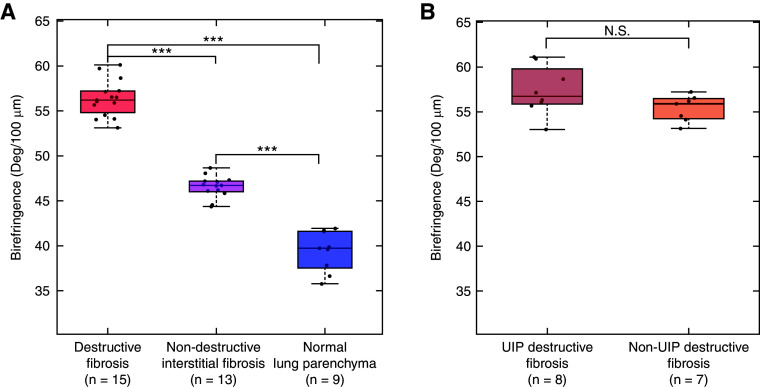
Quantitative measurement of birefringence in fibrosis.
(A) Boxplot of birefringence values from
destructive fibrosis (tissue with destruction of underlying lung
architecture with birefringence present;
n = 15 subjects),
nondestructive interstitial fibrosis (thickening of alveolar walls
with birefringence present;
n = 13 subjects), and normal
lung parenchyma (thin, lattice-like alveoli with birefringence
present; n = 9 subjects).
One-way ANOVA showed statistically significant differences between
mean birefringence across the tissue types for individual subjects
(P < 0.0001). Destructive
fibrosis had the highest mean birefringence, followed by
interstitial fibrosis. Normal lung had the lowest mean
birefringence. Subsequent post hoc Bonferroni tests
confirmed statistically significant differences
(P < 0.0001) between each pair
of tissue types. (B) Further t
test analysis comparing mean birefringence from destructive fibrosis
in histologically confirmed UIP
(n = 8 subjects) versus
destructive fibrosis in non-UIP fILD
(n = 7 subjects) showed no
statistically significant difference
(P = 0.18). In each boxplot,
the data points corresponding to each subject are represented by
dots; the horizontal line in the middle of the box denotes the
median value; the bottom and top edges of the box denote the
25th and 75th percentiles, respectively;
and the whiskers represent the range of data.
***P < 0.0001.
fILD = fibrotic interstitial lung disease;
N.S. = not statistically significant;
UIP = usual interstitial pneumonia.
References
-
- Lynch DA, Sverzellati N, Travis WD, Brown KK, Colby TV, Galvin JR, et al. Diagnostic criteria for idiopathic pulmonary fibrosis: a Fleischner Society white paper. Lancet Respir Med . 2018;6:138–153. - PubMed
-
- Hariri LP, Roden AC, Chung JH, Danoff SK, Gomez Manjarres DC, Hartwig M, et al. The role of surgical lung biopsy in the diagnosis of fibrotic interstitial lung disease: perspective from the Pulmonary Fibrosis Foundation. Ann Am Thorac Soc . 2021;18:1601–1609. - PubMed
Publication types
MeSH terms
Grants and funding
LinkOut - more resources
Full Text Sources
Medical
