. 2022 Jul;41(27):3524-3538.
doi: 10.1038/s41388-022-02362-2. Epub 2022 Jun 9.

RB loss determines selective resistance and novel vulnerabilities in ER-positive breast cancer models

Affiliations

RB loss determines selective resistance and novel vulnerabilities in ER-positive breast cancer models

Vishnu Kumarasamy et al. Oncogene. 2022 Jul.

Abstract

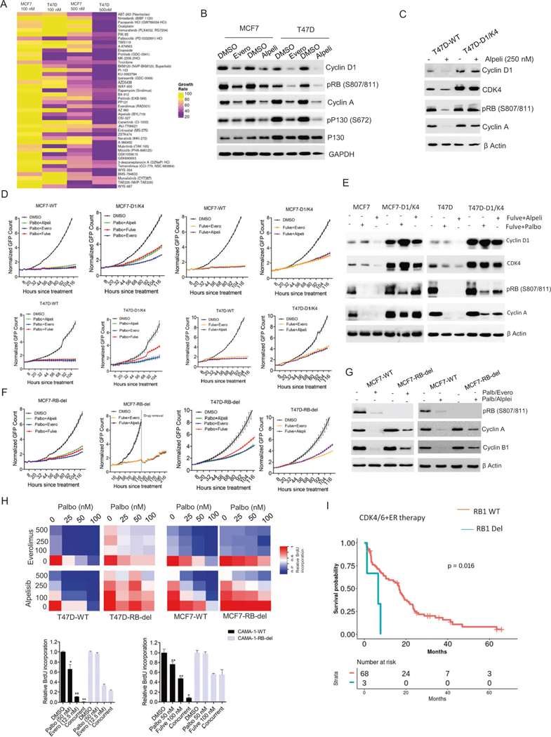
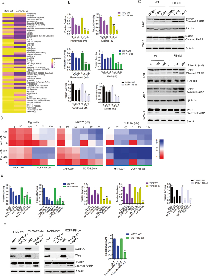
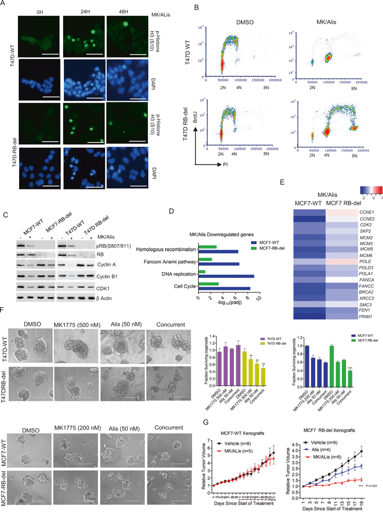
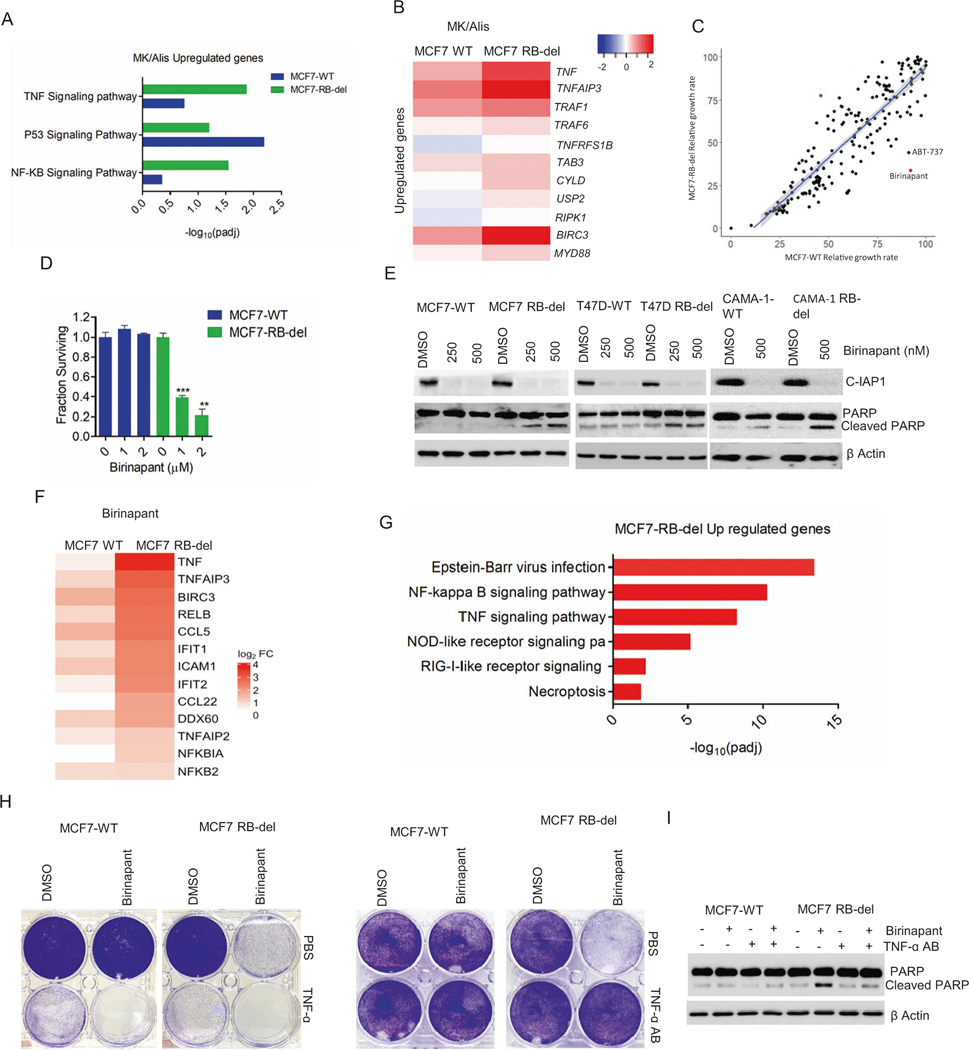
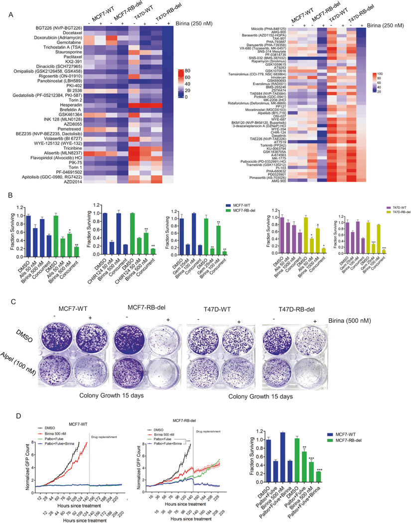
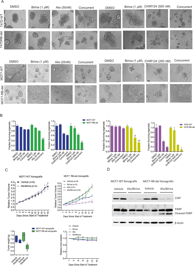
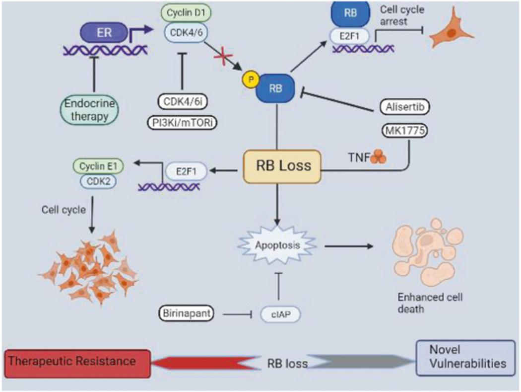
The management of metastatic estrogen receptor (ER) positive HER2 negative breast cancer (ER+) has improved; however, therapeutic resistance and disease progression emerges in majority of cases. Using unbiased approaches, as expected PI3K and MTOR inhibitors emerge as potent inhibitors to delay proliferation of ER+ models harboring PIK3CA mutations. However, the cytostatic efficacy of these drugs is hindered due to marginal impact on the expression of cyclin D1. Different combination approaches involving the inhibition of ER pathway or cell cycle result in durable growth arrest via RB activation and subsequent inhibition of CDK2 activity. However, cell cycle alterations due to RB loss or ectopic CDK4/cyclin D1 activation yields resistance to these cytostatic combination treatments. To define means to counter resistance to targeted therapies imparted with RB loss; complementary drug screens were performed with RB-deleted isogenic cell lines. In this setting, RB loss renders ER+ breast cancer models more vulnerable to drugs that target DNA replication and mitosis. Pairwise combinations using these classes of drugs defines greater selectivity for RB deficiency. The combination of AURK and WEE1 inhibitors, yields synergistic cell death selectively in RB-deleted ER+ breast cancer cells via apoptosis and yields profound disease control in vivo. Through unbiased efforts the XIAP/CIAP inhibitor birinapant was identified as a novel RB-selective agent. Birinapant further enhances the cytotoxic effect of chemotherapies and targeted therapies used in the treatment of ER+ breast cancer models selectively in the RB-deficient setting. Using organoid culture and xenograft models, we demonstrate the highly selective use of birinapant based combinations for the treatment of RB-deficient tumors. Together, these data illustrate the critical role of RB-pathway in response to many agents used to treat ER+ breast cancer, whilst informing new therapeutic approaches that could be deployed against resistant disease.


Conflict of interest statement

ESK and AKW have received research funding from Eli Lilly, Novartis and Pfizer over the last 5 years. There is no current research support from these entities and the study was written in the absence of input from any pharmaceutical company.

Figures

Fig. 1
Fig. 1. RB-mediated cell cycle arrest by standard care of therapies.
A Heat map representing the relative growth rate of MCF7 and T47D cells in the presence of drug library at two different concentrations (100 nM and 500 nM) from cluster 1. B Western blot analysis on the indicated proteins from MCF7 and T47D cells that were treated with everolimus (250 nM) and alpelisib (250 nM) up to 48 H. C Western blot analysis to investigate the effect of alpelisib (250 nM) on cell cycle proteins from T47D and T47D-D1/K4 that were treated up to 48 H. D Live cell imaging to monitor the growth of MCF7-WT, MCF7-D1/K4, T47D-WT, and T47D-D1/K4 cells treated with different pairwise combination of palbociclib (50 nM), fulvestrant (100 nM), everolimus (125 nM) and alpelisib (125 nM). Error bars represent mean and SD from triplicates. Experiments were done at two independent times. E Western blot on the indicated proteins from MCF7-WT, MCF7-D1/K4, T47D-WT and T47D-D1/K4 that were treated with palbociclib (50 nM) + fuvlestrant (100 nM) and alpelisib (125 nM) + fulvestrant (100 nM) for 48 h. F Growth curves illustrating the effect of indicated combination treatments involving palbociclib (50 nM), fulvestrant (100 nM), everolimus (125 nM) and alpelisib (125 nM) in MCF7-RB-del and T47D-RB-del cells. Mean and SD were calculated from triplicates. G Western blot analysis to demonstrate the differential effect of palbociclib (50 nM) + Everolimus (125 nM) and palbociclib (50 nM)+ alpelisib (125 nM) on cell cycle proteins. H Heat map to demonstrate the synergistic effect between the combination of palbociclib with everolimus (Palb/Evero) and alpelisib (Palb/Alpe) on BrdU incorporation at the indicated doses in MCF-WT, MCF7-RB-del, T47D-WT, and T47D-RB-del cells. The values in heat map represent the mean from triplicates. BrdU incorporation was determined in CAMA-1 WT and CAMA-1-RB-del cells treated with the palbociclib (palbo) in combination with everolimus (evero) and fulvestrant (fulve) at indicated concentrations for 72 h. Error bars represent SD from triplicates (*p < 0.05, **p < 0.01 as determined by two-way ANOVA). I PFS of patients receiving CDK4/6i in combination with endocrine therapy with tumors harboring WT RB (n = 68) and RB-deletion mutation (n = 3). All p values were indicated.
Fig. 2
Fig. 2. Vulnerability due to RB loss.
A Heat map representing the differential effect of drugs (250 nM) from cluster 2 on the viability of MCF7-WT and RB-del cells as determined by CTG assay. B Differential effects of alisertib and pemetrexed at the indicated concentrations on the RB-proficient and RB-deficient MCF7, T47D and CAMA-1 cells following 6 days of exposure. Bars represent mean and SD from triplicates. Experiments were performed at 2 independent times (**p < 0.01, ***p < 0.001 as determined by 2-way ANOVA). C Western blot analysis to demonstrate the effect of pemetrexed (Pem) (200 nM) and gemcitabine (Gem) (100 nM) on PARP cleavage in RB-proficient and RB-deficient MCF7 and T47D cells after 72 h treatment. Effect of alisertib at two different concentrations on cleaved PARP in MCF7, T47D and CAMA-1 WT and RB-del cells following 72 h treatment. D Heat maps to demonstrate the synergistic effect on the inhibition of cell viability by different pairwise combination of the indicated drugs in MCF-WT and MCF7-RB-del cells following 6 days of exposure. E CTG assay on MCF7, T47D and CAMA-1 WT/RB-del cell lines following the treatment with the indicated drugs at indicated concentrations as single agents and combination for up to 6 days. Mean and SD were calculated from triplicates (**p < 0.01, ***p < 0.001 as determined by 2-way ANOVA). F Western blot analysis on MCF7-WT, MCF7-RB-del, T47D-WT and T47D-RB-del cells that were reverse transfected with RNAis to silence WEE1 and AURKA. Viability of MCF7-WT and RB-del cells were determined by CTG assay following the knockdowns of WEE1 and AURKA up to 6 days. Bars represent mean and SD from triplicates. Experiments were performed at two independent times (**p < 0.01 as determined by two-way ANOVA). Western blots represent replicates from two independent experiments.
Fig. 3
Fig. 3. Impact of RB loss on AURK and WEE1 inhibition.
A Immunofluorescence staining of p-Histone H3 (S10) on T47D-WT and RB-del cells treated with MK1775 (200 nM) in combination with Alisertib (200 nM) at different time points. Scale bar represents 50 μm. B Representative flow cytometry analysis of T47D-WT and RB-del cells treated with Alisertib (200 nM) and MK1775 (200 nM) for 48 h. X and Y axis represent DNA content and BrdU incorporation respectively. C Western blot analysis on the indicated proteins from RB-proficient and deficient MCF7 and T47D cells that were treated with Alisertib (200 nM) and MK1775 (200 nM) up to 72 H. D ENRICHR analysis to represent the top gene ontology sets that were significantly suppressed in MCF7-WT and MCF7-RB-del cells treated with MK1775 (200 nM) in combination with Alisertib (200 nM) based on RNA sequencing analysis, which was done in triplicates. E Heat map indicating the fold change of selected genes in MCF7WT and RB-deficient cells treated with MK/Alis. F Representative images of organoids derived from T47D-WT, T47D- RB-del, MCF7-WT, and MCF7-RB-del cells that were treated with Alisertib and MK1775 at indicated concentrations for 6 days. The viability of the organoids was determined by CTG assay. Graphs represent mean and SD from triplicates. Experiments were done at two independent times (**p < 0.01, ***p < 0.001 as determined 2-way ANOVA). G Mice bearing MCF7-WT and RB-del xenografts were randomized for treatment with vehicle and MK1775 (30 mg/kg) in combination with Alisertib (10 mg/kg) for 3 weeks and the tumor growth was monitored regularly. Data show mean SEM (***p < 0.001 as determined two-way ANOVA).
Fig. 4
Fig. 4. Regulatory function of RB on apoptosis.
A ENRICHR analysis to represent the top gene ontology sets that were significantly upregulated in MCF7-WT and MCF7-RB-del cells treated with MK/Alis based on RNA sequencing analysis. B Heat map depicting the fold change of indicated genes in MCF7-WT and RB-del cells following the treatment with MK1775 and Alisertib. C Scatter plot with X and Y axis representing the relative growth rate of MCF7-WT and RB-deleted cells respectively that were subjected to drug screen analysis at 500 nM. Each dot represents an individual drug from the library. D CTG assay in MCF7-WT and RB-del cells treated with the increasing concentrations of birinapant up to 6 days. Graphs represent mean and SD from triplicates. Experiments were done at two independent experiments (**p < 0.01, ***p < 0.001 as determined by two-way ANOVA). E Western blot analysis on the indicated proteins from the RB-proficient and RB-deficient cells MCF7, T47D, and CAMA-1 cells treated with different concentrations of birinapant for 72 H. F Heat map representing the fold change of indicated genes in MCF7-WT and RB-del cells following the treatment with birinapant (500 nM) up to 48 h. RNA sequencing was done in triplicates. G ENRICHR analysis to represent the top gene ontology sets that were significantly upregulated in MCF7-WT and MCF7-RB-del cells treated with birinapant (500 nM) based on RNA sequencing analysis. H Representative crystal violet images from MCF7-WT and RB-del cells that were treated with birinapant in the absence and presence of exogenous TNF growth factor (5 ng/μl) and TNF-α antibody (100 ng/μl). I Western blot to determine the effect of birinapant (500 nM) on cleaved PARP in MCF7-WT and RB-del cells in the absence and presence of TNF-α antibody.
Fig. 5
Fig. 5. Combination treatment approach using birinapant.
A Heat maps representing the relative cell viability of MCF7-WT, MCF7-RB-del, T47D-WT and T47D-RB-del cells in the presence of drug library (100 nM) following the pretreatment with birinapant (250 nM) from clusters 1 & 2. B Cell viability assays from MCF7 and T47D WT/RB-del that were treated with birinapant in combination with the indicated drugs at the indicated concentrations up to 6 days. Mean and SD were calculated from triplicates. Experiment was done at 2 independent times. (*p < 0.05, **p < 0.01 as determined by two-way ANOVA). C Crystal violet staining from MCF7-WT, MCF7-RB-del, T47D-WT and T47D-RB-del cells that were treated with Alpelisib (100 nM) in combination with DMSO or birinapant (500 nM). Cells were allowed to form colonies up to 15 days. D Live cell imaging to monitor the proliferation of MCF7-WT and RB-del cells that were treated with DMSO, birinapant (500 nM) and palbociclib (50 nM) + fulvestrant (100 nM) ± birinapant. The live cell imaging was terminated, and the cells were subjected to CTG assay to determine the viable cells. Mean and SD were calculated from 6 replicates. (**p < 0.01, ***p < 0.001 as determined by two-way ANOVA).
Fig. 6
Fig. 6. In vivo efficacy of Alisertib in combination with birinapant.
A Representative images of organoids derived from MCF7-WT, MCF7-RBdel, T47D-WT, and RB-del cells that were treated with birinapant in combination with alisertib and CHIR124 at indicated concentrations for 6 days. B The viability of the organoids were determined by CTG assay. Graphs represent mean and SD from triplicates (*p < 0.05, **p < 0.01, ***p < 0.001 as determined student t-test). C Mice bearing MCF7-WT and RB-del xenografts were randomized for treatment with vehicle, Alisertib (10 mg/kg), birinapant (15 mg/kg) and the combination (Birina/Alis) for 3 weeks and the tumor growth was monitored regularly. Data show mean SEM (***p < 0.001 as determined two-way ANOVA). Box plots represent the tumor weights. Mean and SD are shown (*p < 0.05 as determined by 2-way ANOVA). D Western blot analysis on CIAP1 and cleaved PARP from the MCF7-WT and RB-del tumors at the end of treatment.
Fig. 7
Fig. 7. Scheme illustrating the function of RB loss.
Endocrine therapy, CDK4/6, and PI3K/mTOR inhibitors impact cyclin D1/CDK4/6 function thereby activating RB to induce cell cycle arrest. Loss of RB results in sustained E2F activity even in the presence of cytostatic drugs that transactivates cyclin E to drive cell cycle via CDK2 activity. Alisertib in combination with MK1775 induces apoptosis selectively in RB-deficient setting. Birinapant, an inhibitor of IAP, which is an anti-apoptotic protein results in enhanced apoptotic mediated cell death in RB -deficient ER + breast cancer models. Overall, RB loss leads to therapeutic resistance to the standard cytostatic treatment options and becomes more vulnerable to pharmacological agents that induce apoptosis.

Similar articles

Cited by

References

    1. Gong Y, Liu YR, Ji P, Hu X, Shao ZM. Impact of molecular subtypes on metastatic breast cancer patients: a SEER population-based study. Sci Rep. 2017;7:45411. - PMC - PubMed
    1. Ahmad A. Breast cancer statistics: recent trends. Adv Exp Med Biol. 2019;1152:1–7. - PubMed
    1. Jensen EV, Jordan VC. The estrogen receptor: a model for molecular medicine. Clin Cancer Res. 2003;9:1980–9. - PubMed
    1. Howell A. The endocrine prevention of breast cancer. Best Pract Res Clin Endocrinol Metab. 2008;22:615–23. - PubMed
    1. Mouridsen H, Gershanovich M, Sun Y, Perez-Carrion R, Boni C, Monnier A, et al. Superior efficacy of letrozole versus tamoxifen as first-line therapy for postmenopausal women with advanced breast cancer: results of a phase III study of the International Letrozole Breast Cancer Group. J Clin Oncol. 2001;19:2596–606. - PubMed

Publication types