Tubo-ovarian mass with raised CA-125 in a 21-year-old female
- PMID: 35676687
- PMCID: PMC9178888
- DOI: 10.1186/s12957-022-02651-w
Tubo-ovarian mass with raised CA-125 in a 21-year-old female
Abstract
Introduction: Peritonitis associated with fungal species Curvularia lunata seldom occurs with only five cases reported in the literature, all in middle-age patients with comorbidities undergoing dialysis.
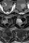
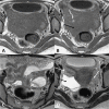
Case report: A 21-year-old female who was referred to surgical oncology OPD with a diagnosis of ovarian malignancy, based on raised cancer antigen 125 (CA 125) and suspected tubo-ovarian mass (TOM) on magnetic resonance imaging (MRI). A review of the MRI showed a pelvic collection with TOM, suggestive of infective pathology. Fungal culture and mass spectroscopy of the cystic collection identified the presence of Curvularia lunata. She was treated with oral itraconazole which showed symptomatic improvement and radiological response. In the follow-up period, the patient developed chest wall swelling, aspiration and geneXpert® revealed multidrug-resistant (MDR) tuberculosis, and treatment was started.
Conclusions: Unusual causes of TOM and raised CA 125 should be kept in mind when dealing with young patients, as the possibility of epithelial ovarian cancer in this age is very low.
Keywords: Curvularia; Fungal; Infection; Peritonitis; Young.
© 2022. The Author(s).
Conflict of interest statement
The authors declare that they have no competing interests.
Figures


Similar articles
-
Curvularia geniculata fungal peritonitis: a case report with review of literature.Int Urol Nephrol. 2005;37(4):781-4. doi: 10.1007/s11255-004-0628-4. Int Urol Nephrol. 2005. PMID: 16362599 Review.
-
Curvularia lunata, a rare fungal peritonitis in continuous ambulatory peritoneal dialysis (CAPD); a rare case report.J Nephropharmacol. 2015 Dec 21;5(1):61-62. eCollection 2016. J Nephropharmacol. 2015. PMID: 28197501 Free PMC article.
-
Severe influenza A virus pneumonia complicated with Curvularia lunata infection: Case Report.Front Cell Infect Microbiol. 2023 Dec 15;13:1289235. doi: 10.3389/fcimb.2023.1289235. eCollection 2023. Front Cell Infect Microbiol. 2023. PMID: 38162579 Free PMC article.
-
Peritonitis due to Curvularia inaequalis in an elderly patient undergoing peritoneal dialysis and a review of six cases of peritonitis associated with other Curvularia spp.J Clin Microbiol. 2005 Aug;43(8):4288-92. doi: 10.1128/JCM.43.8.4288-4292.2005. J Clin Microbiol. 2005. PMID: 16082004 Free PMC article. Review.
-
Pelvic mass associated with raised CA 125 for benign condition: a case report.World J Surg Oncol. 2010 Apr 16;8:28. doi: 10.1186/1477-7819-8-28. World J Surg Oncol. 2010. PMID: 20398372 Free PMC article.
Cited by
-
Epidemiology of rare bacterial, parasitic, and fungal pathogens in India.IJID Reg. 2024 Mar 21;11:100359. doi: 10.1016/j.ijregi.2024.100359. eCollection 2024 Jun. IJID Reg. 2024. PMID: 38646508 Free PMC article. Review.
-
Massive serous cyst adenoma with ovarian abscess causing fatal septicaemia.BMJ Case Rep. 2023 Nov 20;16(11):e255467. doi: 10.1136/bcr-2023-255467. BMJ Case Rep. 2023. PMID: 37989331
-
Sensitivity of Immunodiagnostic Tests in Localized Versus Disseminated Tuberculosis-A Systematic Review of Individual Patient Data.Trop Med Infect Dis. 2025 Mar 7;10(3):70. doi: 10.3390/tropicalmed10030070. Trop Med Infect Dis. 2025. PMID: 40137824 Free PMC article. Review.
References
Publication types
MeSH terms
Substances
Supplementary concepts
LinkOut - more resources
Full Text Sources
Medical
Research Materials
Miscellaneous

