

Elevated Patellofemoral and Tibiofemoral T1ρ Relaxation Times Following a First Time Patellar Dislocation
- PMID: 35676874
- PMCID: PMC9189536
- DOI: 10.1177/19476035221102570
Elevated Patellofemoral and Tibiofemoral T1ρ Relaxation Times Following a First Time Patellar Dislocation
Abstract
Objective: The study was performed to evaluate cartilage within the knee following a first-time patellar dislocation, using elevated MRI-based T1ρ relaxation times as an indicator of low proteoglycan concentration. The hypothesis is that MRI-based T1ρ relaxation times for patellofemoral and tibiofemoral cartilage are significantly longer for knees being treated for patellar dislocation than for healthy control knees.
Design: Twenty-one subjects being treated for a first-time, unilateral dislocation of the patella and 16 healthy controls participated in MRI-based T1ρ relaxation time mapping. Mean relaxation times were quantified for patellofemoral and tibiofemoral regions for injured knees, the contralateral knees, and healthy controls. T1ρ values for each region were compared between the 3 groups with generalized estimating equations. Linear regressions were also performed to correlate T1ρ relaxation times with time from injury.
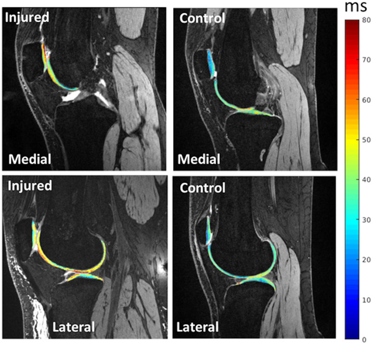
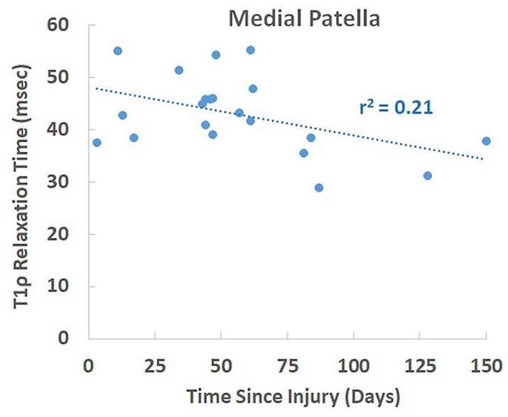
Results: The knees with a disloction had longer T1ρ relaxation times than the contralateral knees and control group at the medial patella and longer relaxation times than the control group at the lateral tibia (P < 0.05). T1ρ relaxation times at the medial patella also decreased with time from injury (r2 = 0.21, P = 0.037).
Conclusions: Compositional changes to cartilage on the medial patella are related to traumatic impact during a dislocation. Potential exists for cartilage properties at the medial patella to improve with time. Cartilage degradation at the lateral tibia is not directly related to traumatic impact. The current baseline data are a starting point to characterize the pathway from a first-time dislocation to progressive cartilage degradation and osteoarthritis.
Keywords: T1ρ; cartilage; patellar dislocation.
Conflict of interest statement
Figures
Similar articles
-
Patellar Dislocation in Adolescent Patients: Influence on Cartilage Properties Based on T1ρ Relaxation Times.Am J Sports Med. 2023 Dec;51(14):3714-3723. doi: 10.1177/03635465231205562. Epub 2023 Oct 28. Am J Sports Med. 2023. PMID: 37897349 Free PMC article.
-
Patellofemoral Cartilage Degradation Based on T1ρ Relaxation Times Varies Inversely With BMI After Patellar Dislocation.Orthop J Sports Med. 2025 May 20;13(5):23259671251334634. doi: 10.1177/23259671251334634. eCollection 2025 May. Orthop J Sports Med. 2025. PMID: 40401089 Free PMC article.
-
In Vivo Compositional Changes in the Articular Cartilage of the Patellofemoral Joint Following Anterior Cruciate Ligament Reconstruction.Arthritis Care Res (Hoboken). 2022 Jul;74(7):1172-1178. doi: 10.1002/acr.24561. Epub 2022 Apr 13. Arthritis Care Res (Hoboken). 2022. PMID: 33460530 Free PMC article.
-
Chondral and osteochondral injuries associated with acute patellar dislocation.Arthroscopy. 2003 Sep;19(7):717-21. doi: 10.1016/s0749-8063(03)00401-8. Arthroscopy. 2003. PMID: 12966379 Review.
-
Intra-articular horizontal dislocation of the patella: a rare injury and review of the literature.BMJ Case Rep. 2020 Jul 9;13(7):e232249. doi: 10.1136/bcr-2019-232249. BMJ Case Rep. 2020. PMID: 32646932 Free PMC article. Review.
Cited by
-
Patellar Dislocation in Adolescent Patients: Influence on Cartilage Properties Based on T1ρ Relaxation Times.Am J Sports Med. 2023 Dec;51(14):3714-3723. doi: 10.1177/03635465231205562. Epub 2023 Oct 28. Am J Sports Med. 2023. PMID: 37897349 Free PMC article.
-
Adding Tibial Tuberosity Medialization to Medial Patellofemoral Ligament Reconstruction Reduces Lateral Patellar Maltracking During Multidirectional Motion in a Computational Simulation Model.Arthrosc Sports Med Rehabil. 2023 Jul 10;5(4):100753. doi: 10.1016/j.asmr.2023.100753. eCollection 2023 Aug. Arthrosc Sports Med Rehabil. 2023. PMID: 37645404 Free PMC article.
-
Patellofemoral Cartilage Degradation Based on T1ρ Relaxation Times Varies Inversely With BMI After Patellar Dislocation.Orthop J Sports Med. 2025 May 20;13(5):23259671251334634. doi: 10.1177/23259671251334634. eCollection 2025 May. Orthop J Sports Med. 2025. PMID: 40401089 Free PMC article.
References
Publication types
MeSH terms
LinkOut - more resources
Full Text Sources
Medical
