. 2022 May 23:13:896179.
doi: 10.3389/fimmu.2022.896179. eCollection 2022.

A Holistic View of the Goto-Kakizaki Rat Immune System: Decreased Circulating Immune Markers in Non- Obese Type 2 Diabetes

Affiliations

A Holistic View of the Goto-Kakizaki Rat Immune System: Decreased Circulating Immune Markers in Non- Obese Type 2 Diabetes

Snehaa V Seal et al. Front Immunol. .

Abstract

Type-2 diabetes is a complex disorder that is now considered to have an immune component, with functional impairments in many immune cell types. Type-2 diabetes is often accompanied by comorbid obesity, which is associated with low grade inflammation. However,the immune status in Type-2 diabetes independent of obesity remains unclear. Goto-Kakizaki rats are a non-obese Type-2 diabetes model. The limited evidence available suggests that Goto-Kakizaki rats have a pro-inflammatory immune profile in pancreatic islets. Here we present a detailed overview of the adult Goto-Kakizaki rat immune system. Three converging lines of evidence: fewer pro-inflammatory cells, lower levels of circulating pro-inflammatory cytokines, and a clear downregulation of pro-inflammatory signalling in liver, muscle and adipose tissues indicate a limited pro-inflammatory baseline immune profile outside the pancreas. As Type-2 diabetes is frequently associated with obesity and adipocyte-released inflammatory mediators, the pro-inflammatory milieu seems not due to Type-2 diabetes per se; although this overall reduction of immune markers suggests marked immune dysfunction in Goto-Kakizaki rats.

Keywords: Goto-Kakizaki rats; cytokines; diabetes; inflammation; microarrays; obesity.


Conflict of interest statement

The authors declare that the research was conducted in the absence of any commercial or financial relationships that could be construed as a potential conflict of interest.

Figures

Figure 1
Figure 1
The diabetic milieu majorly alters the GK immune system. Significantly affected immune cells (p<0.05), expressed as percentage of parent population frequency are, (A) CD11b/c+ phagocytes (macrophages, monocytes, dendritic cells, granulocytes), (B) CD45RA+ B cells, (C) CD4+ T-helper cells, (D) CD8+ T-cytotoxic cells (E) CD161a+ NK cells, T-cell subsets, activated monocytes, and dendritic cells (F) CD8+ RORγT+ Tc17 cells (G) GATA3+ Tc2 cells. However, the T-helper cells narrowly missed the significance threshold (p = 0.0649). Data are mean +/- SEM; individual animals shown as dots. Grey bars: GK rats (n = 6); white bars, Wistar rats. (n = 6).
Figure 2
Figure 2
Overall cytokine profile shows limited peripheral inflammation in GK rats. All panels display the mean levels of plasma cytokines (mean +/- SEM) in pg/mL, that were significantly different (p<0.05) in the GK rats compared to the controls. Cytokines shown in panels (A–M) are proinflammatory, (N) is both pro- and anti-inflammatory, (O–Q) are anti-inflammatory while (R, S) show no inflammatory role. Grey bars: GK rats (n = 7); white bars: Wistar rats (n = 6). p < 0.001, p < 0.01 and p < 0.05 are reflected by ***, ** and * respectively. Student’s t-test was used except for panels E (IL-2), L (LIX), and R (IL5), which were analysed using a Mann-Whitney test.
Figure 3
Figure 3
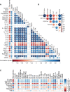
Integrative correlation analysis of the different immune parameters, (A) Cytokine-cytokine (B) Immune cell-immune cell (C) Cytokine-immune cells. The sizes of the circles indicate the strength of association (Pearson’s r2), while the colours show negative (red) or positive (blue) correlation. p < 0.001, p < 0.01 and p < 0.05 are reflected by ***, ** and * respectively.
Figure 4
Figure 4
Transcriptomic re-analysis of 3 principal diabetic tissues (liver, muscle and adipose tissue) in Wistar rats compared to GK rats. (A) Venn diagram showing the number of common biological pathways. (B) Biological pathways that are regulated by downstream signalling from circulating cytokines analysed in the muscle. (C) Biological pathways associated with diabetes and its complications analysed in the muscle. For (B, C), the intensity of the colour is proportional to the -log10 of P-value, sizes of the circles represent the number of genes involved, while placement of the circles on the x axis indicates the fold enrichment. (D–G) Are T-cell receptor signalling, Jak-STAT signalling, TGF-β signalling and PDL1 signalling respectively in liver, muscle and adipose tissue. Data from GSE13271 (16).

References

    1. Berbudi A, Rahmadika N, Tjahjadi AI, Ruslami R. Type 2 Diabetes and its Impact on the Immune System. Curr Diabetes Rev (2020) 16(5):442–9. doi: 10.2174/1573399815666191024085838 - DOI - PMC - PubMed
    1. de Candia P, Prattichizzo F, Garavelli S, De Rosa V, Galgani M, Di Rella F, et al. . Type 2 Diabetes: How Much of an Autoimmune Disease? Front Endocrinol (Lausanne) (2019) 10:451. doi: 10.3389/fendo.2019.00451 - DOI - PMC - PubMed
    1. Ahlqvist E, Prasad RB, Groop L. Subtypes of Type 2 Diabetes Determined From Clinical Parameters. Diabetes (2020) 69(10):2086–93. doi: 10.2337/dbi20-0001 - DOI - PubMed
    1. Kietsiriroje N, Pearson S, Campbell M, Ariens RAS, Ajjan RA. Double Diabetes: A Distinct High-Risk Group? Diabetes Obes Metab (2019) 21(12):2609–18. doi: 10.1111/dom.13848 - DOI - PubMed
    1. Takeda Y, Shimomura T, Asao H, Wakabayashi I. Relationship Between Immunological Abnormalities in Rat Models of Diabetes Mellitus and the Amplification Circuits for Diabetes. J Diabetes Res (2017) 2017:4275851. doi: 10.1155/2017/4275851 - DOI - PMC - PubMed

Publication types