[Preprint]. 2022 Jun 1:2022.05.31.494211.
doi: 10.1101/2022.05.31.494211.

SARS-CoV-2 Variant Spike and accessory gene mutations alter pathogenesis

Affiliations

SARS-CoV-2 Variant Spike and accessory gene mutations alter pathogenesis

M E McGrath et al. bioRxiv. .

Update in

  • SARS-CoV-2 variant spike and accessory gene mutations alter pathogenesis.
    McGrath ME, Xue Y, Dillen C, Oldfield L, Assad-Garcia N, Zaveri J, Singh N, Baracco L, Taylor LJ, Vashee S, Frieman MB. McGrath ME, et al. Proc Natl Acad Sci U S A. 2022 Sep 13;119(37):e2204717119. doi: 10.1073/pnas.2204717119. Epub 2022 Aug 30. Proc Natl Acad Sci U S A. 2022. PMID: 36040867 Free PMC article.

Abstract

The ongoing COVID-19 pandemic is a major public health crisis. Despite the development and deployment of vaccines against SARS-CoV-2, the pandemic persists. The continued spread of the virus is largely driven by the emergence of viral variants, which can evade the current vaccines through mutations in the Spike protein. Although these differences in Spike are important in terms of transmission and vaccine responses, these variants possess mutations in the other parts of their genome which may affect pathogenesis. Of particular interest to us are the mutations present in the accessory genes, which have been shown to contribute to pathogenesis in the host through innate immune signaling, among other effects on host machinery. To examine the effects of accessory protein mutations and other non-spike mutations on SARS-CoV-2 pathogenesis, we synthesized viruses where the WA1 Spike is replaced by each variant spike genes in a SARS-CoV-2/WA-1 infectious clone. We then characterized the in vitro and in vivo replication of these viruses and compared them to the full variant viruses. Our work has revealed that non-spike mutations in variants can contribute to replication of SARS-CoV-2 and pathogenesis in the host and can lead to attenuating phenotypes in circulating variants of concern. This work suggests that while Spike mutations may enhance receptor binding and entry into cells, mutations in accessory proteins may lead to less clinical disease, extended time toward knowing an infection exists in a person and thus increased time for transmission to occur.

Significance: A hallmark of the COVID19 pandemic has been the emergence of SARS-CoV-2 variants that have increased transmission and immune evasion. Each variant has a set of mutations that can be tracked by sequencing but little is known about their affect on pathogenesis. In this work we first identify accessory genes that are responsible for pathogenesis in vivo as well as identify the role of variant spike genes on replication and disease in mice. Isolating the role of Spike mutations in variants identifies the non-Spike mutations as key drivers of disease for each variant leading to the hypothesis that viral fitness depends on balancing increased Spike binding and immuno-evasion with attenuating phenotypes in other genes in the SARS-CoV-2 genome.


Figures

Figure 1.
Figure 1.. Assembly of infectious clone genomes of SARS-CoV-2.
A. The genome of WA1 was assembled from sequence-validated overlapping (colored ends) DNA fragments (1a-1, 1a-2, 1a-3, 1b-1, 1b-2, S, AP; blue lines) by TAR in yeast. The infectious clone genomes can be maintained in yeast and E. coli by a YCpBAC vector. The infectious clone genome, which is flanked by I-SceI sites, is driven by a CMV promoter (PCMV) and has a hepatitis delta virus ribozyme sequence (Hdz) as well as a bovine growth hormone terminator (Term) at the 3’ end of the genome. SARS-CoV-2 WA1 genomes containing either Spike variants or accessory ORF deletions were assembled from a mix of unmodified and appropriate modified DNA fragments. B. PCR amplifications of assembly junctions (J1–10) to confirm a full-length genome in E. coli. M, 2-log marker.
Figure 2.
Figure 2.. WA-1 accessory deletion viruses in 12-week old K18-hACE2 mice.
A. Supernatant titers of VeroE6 cells infected with an M.O.I. of 0.01 of accessory deletion viruses with supernatant pulled at 0, 6, 24, 48, 72, and 96 hours and titered by plaque assay. B. Percent starting weight on days 0–4 of K18-hACE2 mice infected with 1e3 pfu of each accessory deletion virus. C. Lung viral titers of mice euthanized on D2 and D4 by pfu/g lung. D. Brain viral titers of mice euthanized on D2 and D4 by pfu/g brain. E. Lung viral loads of mice euthanized on D2 and D4 by qPCR for Rdrp. F. Brain viral loads of mice euthanized on D2 and D4 by qPCR for Rdrp. (*= pfu ≤ 0.05, **=pfu ≤ 0.005, ***= pfu ≤ 0.0005)
Figure 3.
Figure 3.. Lung pathology of 12-week old K18-hACE2 mice infected with accessory deletion viruses of WA-1.
A. H/E stained sections of the lungs. B. Pathological scoring of the lungs.
Figure 4.
Figure 4.. Variant spikes in WA-1 viruses in 12-week old K18-hACE2 mice.
A. Supernatant titers of VeroE6 cells infected with an M.O.I. of 0.01 of variant spikes in WA-1 and parent variant viruses with supernatant pulled at 0, 6, 24, 48. 72, and 96 hours and titered by plaque assay. B. Percent starting weight on days 0–4 of K18-hACE2 mice infected with 1e3 pfu of each variant spike in WA-1 virus or the parent variant virus. C. Lung viral titers of mice euthanized on D2 and D4 by pfu/g lung. D. Brain viral titers of mice euthanized on D2 and D4 by pfu/g brain. E. Lung viral loads of mice euthanized on D2 and D4 by qPCR for Rdrp. F. Brain viral loads of mice euthanized on D2 and D4 by qPCR for Rdrp.
Figure 5.
Figure 5.. Lung pathology of 12-week old K18-hACE2 mice infected with variant spikes in WA-1 and parent variants.
A. H/E stained lung sections. B. Pathological scoring of the lungs.
Figure 6.
Figure 6.. Variant spikes in WA-1 viruses in 12-week old BALB/c mice.
A. Percent starting weight on day 0–4 of BALB/c mice infected with 1e4 pfu of each variant spike in WA-1 virus. B. Lung viral titers of mice euthanized on D2 and D4 by pfu/g lung. C. Lung viral titers of mice euthanized on D2 and D4 by qPCR for Rdrp.
Figure 7.
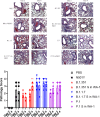
Figure 7.. Lung pathology of 12-week old BALB/c mice infected with variant spike in WA-1 viruses and parent variants.
A. H/E stained lung sections. B. Pathological scoring of the lungs.

References

    1. Zhou P. et al. A pneumonia outbreak associated with a new coronavirus of probable bat origin. Nature 579, 270–273 (2020). - PMC - PubMed
    1. Archived: WHO Timeline - COVID-19. https://www.who.int/news/item/27-04-2020-who-timeline--covid-19.
    1. WHO Coronavirus (COVID-19) Dashboard. https://covid19.who.int.
    1. Weston S. & Frieman M. B. COVID-19: Knowns, Unknowns, and Questions. mSphere (2020) doi:10.1128/mSphere.00203-20. - DOI - PMC - PubMed
    1. Hu B., Guo H., Zhou P. & Shi Z.-L. Characteristics of SARS-CoV-2 and COVID-19. Nat. Rev. Microbiol. 19, 141–154 (2021). - PMC - PubMed

Publication types