Review
. 2022 May 23:10:889431.
doi: 10.3389/fbioe.2022.889431. eCollection 2022.

Recent Advances in Aptasensors For Rapid and Sensitive Detection of Staphylococcus Aureus

Affiliations
Review

Recent Advances in Aptasensors For Rapid and Sensitive Detection of Staphylococcus Aureus

Wei Chen et al. Front Bioeng Biotechnol. .

Abstract

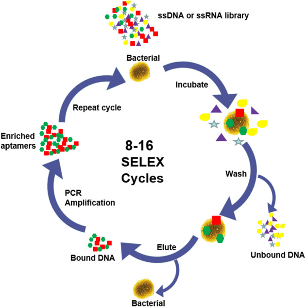
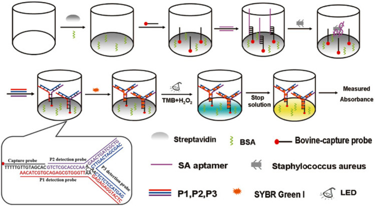
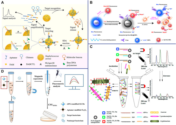
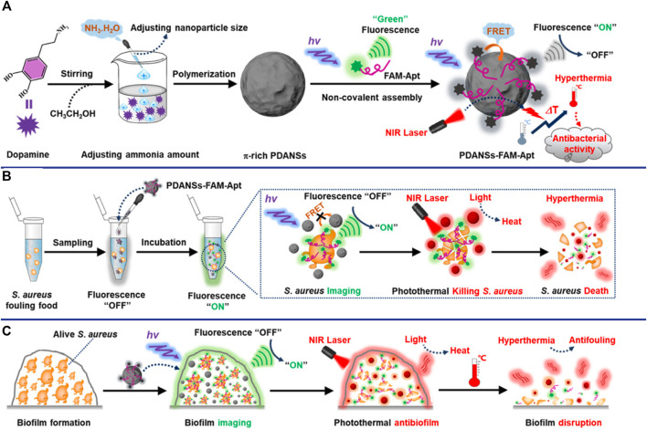
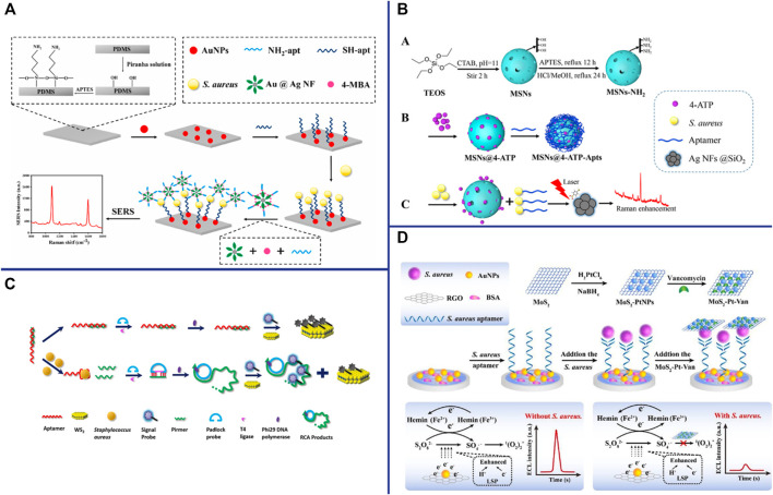
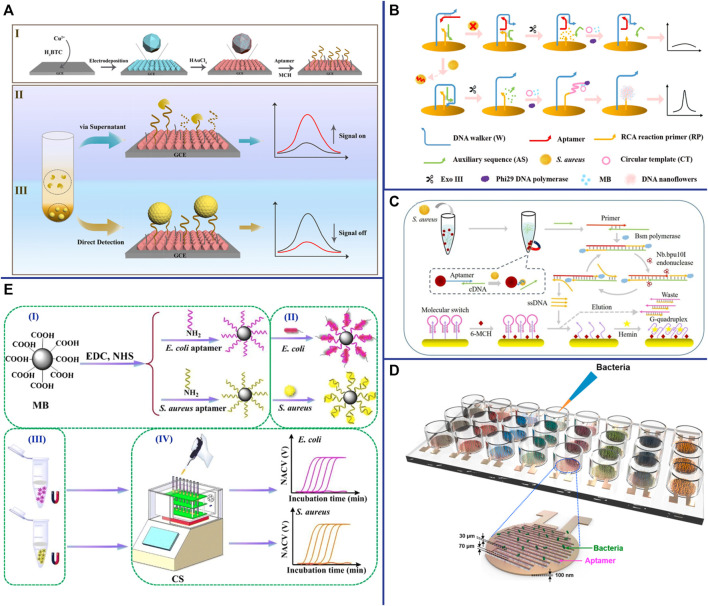
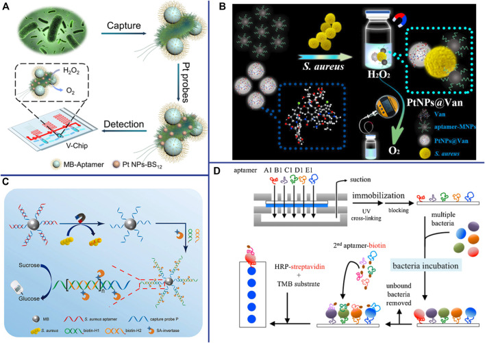
The infection of Staphylococcus aureus (S.aureus) and the spread of drug-resistant bacteria pose a serious threat to global public health. Therefore, timely, rapid and accurate detection of S. aureus is of great significance for food safety, environmental monitoring, clinical diagnosis and treatment, and prevention of drug-resistant bacteria dissemination. Traditional S. aureus detection methods such as culture identification, ELISA, PCR, MALDI-TOF-MS and sequencing, etc., have good sensitivity and specificity, but they are complex to operate, requiring professionals and expensive and complex machines. Therefore, it is still challenging to develop a fast, simple, low-cost, specific and sensitive S. aureus detection method. Recent studies have demonstrated that fast, specific, low-cost, low sample volume, automated, and portable aptasensors have been widely used for S. aureus detection and have been proposed as the most attractive alternatives to their traditional detection methods. In this review, recent advances of aptasensors based on different transducer (optical and electrochemical) for S. aureus detection have been discussed in details. Furthermore, the applications of aptasensors in point-of-care testing (POCT) have also been discussed. More and more aptasensors are combined with nanomaterials as efficient transducers and amplifiers, which appears to be the development trend in aptasensors. Finally, some significant challenges for the development and application of aptasensors are outlined.

Keywords: POCT; Staphylococcus aureus; aptasensor; electrochemical biosensor; nanomaterials; optical biosensor.


Conflict of interest statement

The authors declare that the research was conducted in the absence of any commercial or financial relationships that could be construed as a potential conflict of interest.

Figures

FIGURE 1
FIGURE 1
SELEX process.
FIGURE 2
FIGURE 2
Schematic illustration of the principle for detection of S. aureus with aptamer-high throughput colorimetric biosensor based on photocatalytic activity of dsDNA-SG I complex. Reproduced with permission (Yu et al., 2020).
FIGURE 3
FIGURE 3
(A) Schematic representation of the fluorescent detection of S. aureus based on a finely designed functional chimera sequence, a molecular beacon (MB), and strand displacement target recycling. Reproduced with permission (Cai et al., 2019). (B) General design of the vancomycin and aptamer dual-recognition moieties-based ratiometric fluorescent nanoprobe with a remarkably large Stokes shift for ultrafast and accurate management of S. aureus at the single-cell level. Reproduced with permission (Shen et al., 2020). (C) Schematic illustration of the multiplexed luminescence bioassay based on aptamers-modified UCNPs for the simultaneous detection of various pathogenic bacteria. Reproduced with permission (Wu et al., 2014). (D) Schematic illustration of the fluorometric aptasensor for high-sensitivity S. aureus detection based on FRET of the self-assembled dimer, B-CD/Fe3O4. Reproduced with permission (Cui et al., 2019).
FIGURE 4
FIGURE 4
General design of the smart nanoprobe PDANSs-FAM-Apt for accurate fluorescence detection and imaging-guided precise photothermal antibacteria. (A) Illustration of the assembly procedure of the nanoprobe PDANSs-FAM-Apt. (B) Diagram of the FRET-based assay procedure for PDANSs-FAM-Apt responsive to living S. aureus and imaging-guided photothermal killing of S. aureus. (C) Schematic of imaging-guided photothermal antifouling of the nanoprobe PDANSs-FAM-Apt for the destruction of S. aureus biofilms. Reproduced with permission (Ye et al., 2020).
FIGURE 5
FIGURE 5
(A) Schematic illustration of the developed SERS biosensor based on aptamer functionalized PDMS film for the detection of S. aureus. Reproduced with permission (Zhu et al., 2021a). (B) Schematic illustration of S. aureus detection based on the target-responsive release of 4-ATP molecules from aptamer-gated MSNs. Reproduced with permission (Zhu et al., 2021b). (C) Schematic illustration of the CRET biosensor for the detection of S. aureus based on Co2+/ABEI-AuNFs and WS2 nanosheet. Reproduced with permission (Hao et al., 2017). (D) Schematic illustration of the enzyme-free ECL aptasensor for S. aureus detection based on AuNPs/hemin as the regenerable enhancers of S2O8 2−/O2 and the quenching effect of MoS2-PtNPs on S2O8 2−/O2. Reproduced with permission (Han et al., 2019).
FIGURE 6
FIGURE 6
(A) (I) Fabrication of the DNA/AuNPs/MOFs-based sensing platform; (II) Schematic diagrams of the electrochemical biosensor for the detection of S. aureus via supernatants; and (III) pathogen cells. Reproduced with permission (Sun et al., 2021). (B) Schematic representation of the biosensor for S. aureus detection based on a DNA walker and DNA nanoflower. Reproduced with permission (Cai et al., 2021b). (C) Schematic representation of the versatile signal-on electrochemical biosensor for S. aureus detection based on triple-helix molecular switch. Reproduced with permission (Cai et al., 2021a). (D) Schematic of aptamer-functionalized capacitance sensor array for real-time monitoring of bacterial growth and antibiotic susceptibility. Reproduced with permission (Jo et al., 2018). (E) Schematic representation of the multichannel conductometric sensor for S. aureus detection based on magnetic analyte separation via aptamer. (I)the preparation of aptamer-functionalized magnetic beads, (II) selective capture and (III) separation of bacterial cells, and (IV) determination of viable bacteria by the conductometric sensor. Reproduced with permission (Zhang et al., 2020).
FIGURE 7
FIGURE 7
(A) Working principle for detection of bacteria using BV-Chip. Reproduced with permission (Huang et al., 2019). (B) Schematic illustration of gas pressure-based POC testing protocol for highly sensitive and specific detection of S. aureus. Reproduced with permission (Li J. et al., 2019a). (C) Schematic illustration of the principle for portable detection of S. aureus using PGM based on HCR strategy. Reproduced with permission (Yang et al., 2021b). (D) The experimental procedure for multi-bacterial detection via bacteria-specific aptamers immobilized on a nitrocellulose (NC) membrane. 2nd (secondary) aptamer-biotin conjugated with biotin. Reproduced with permission (Wang et al., 2019a).

Similar articles

Cited by

References

    1. Abbaspour A., Norouz-Sarvestani F., Noori A., Soltani N. (2015). Aptamer-conjugated Silver Nanoparticles for Electrochemical Dual-Aptamer-Based Sandwich Detection of staphylococcus Aureus. Biosens. Bioelectron. 68, 149–155. 10.1016/j.bios.2014.12.040 - DOI - PubMed
    1. Alizadeh N., Memar M. Y., Moaddab S. R., Kafil H. S. (2017). Aptamer-assisted Novel Technologies for Detecting Bacterial Pathogens. Biomed. Pharmacother. 93, 737–745. 10.1016/j.biopha.2017.07.011 - DOI - PubMed
    1. Amraee M., Oloomi M., Yavari A., Bouzari S. (2017). DNA Aptamer Identification and Characterization for E. coli O157 Detection Using Cell Based SELEX Method. Anal. Biochem. 536, 36–44. 10.1016/j.ab.2017.08.005 - DOI - PubMed
    1. Banerjee J. (2010). Antibodies Are Challenged. Indian J. Med. Sci. 64, 144–147. - PubMed
    1. Berezovski M., Musheev M., Drabovich A., Krylov S. N. (2006a). Non-SELEX Selection of Aptamers. J. Am. Chem. Soc. 128, 1410–1411. 10.1021/ja056943j - DOI - PubMed

LinkOut - more resources