. 2022 May 23:13:884449.
doi: 10.3389/fneur.2022.884449. eCollection 2022.

Cerebral Microbleeds Assessment and Quantification in COVID-19 Patients With Neurological Manifestations

Affiliations

Cerebral Microbleeds Assessment and Quantification in COVID-19 Patients With Neurological Manifestations

Angela Napolitano et al. Front Neurol. .

Abstract

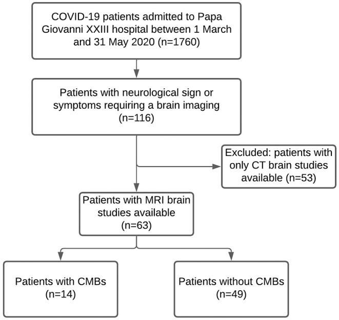
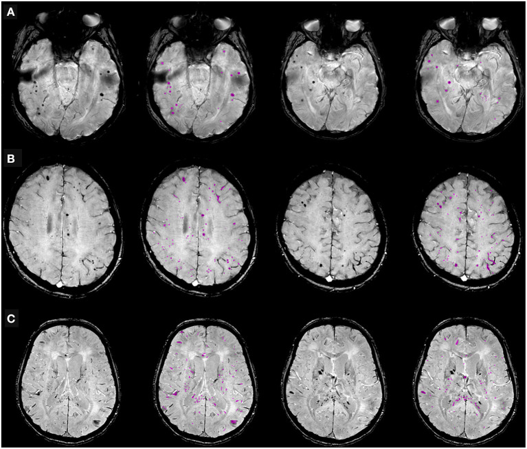
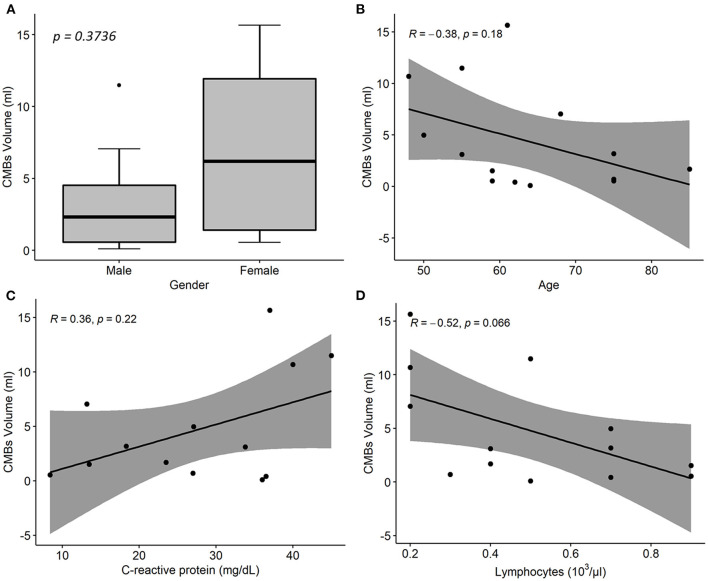
It is increasingly acknowledged that Coronavirus Disease 2019 (COVID-19) can have neurological manifestations, and cerebral microbleeds (CMBs) have been observed in this setting. The aim of this study was to characterize CMBs patterns on susceptibility-weighted imaging (SWI) in hospitalized patients with COVID-19 with neurological manifestations. CMBs volume was quantified and correlated with clinical and laboratory parameters. The study included patients who were hospitalized due to COVID-19, exhibited neurological manifestations, and underwent a brain MRI between March and May 2020. Neurological, clinical, and biochemical variables were reported. The MRI was acquired using a 3T scanner, with a standardized protocol including SWI. Patients were divided based on radiological evidence of CMBs or their absence. The CMBs burden was also assessed with a semi-automatic SWI processing procedure specifically developed for the purpose of this study. Odds ratios (OR) for CMBs were calculated using age, sex, clinical, and laboratory data by logistic regression analysis. Of the 1,760 patients with COVID-19 admitted to the ASST Papa Giovanni XXIII Hospital between 1 March and 31 May 2020, 116 exhibited neurological symptoms requiring neuroimaging evaluation. Of these, 63 patients underwent brain MRI and were therefore included in the study. A total of 14 patients had radiological evidence of CMBs (CMBs+ group). CMBs+ patients had a higher prevalence of CSF inflammation (p = 0.020), a higher white blood cell count (p = 0.020), and lower lymphocytes (p = 0.010); the D-dimer (p = 0.026), LDH (p = 0.004), procalcitonin (p = 0.002), and CRP concentration (p < 0.001) were higher than in the CMBs- group. In multivariable logistic regression analysis, CRP (OR = 1.16, p = 0.011) indicated an association with CMBs. Estimated CMBs volume was higher in females than in males and decreased with age (Rho = -0.38; p = 0.18); it was positively associated with CRP (Rho = 0.36; p = 0.22), and negatively associated with lymphocytes (Rho = -0.52; p = 0.07). CMBs are a frequent imaging finding in hospitalized patients with COVID-19 with neurological manifestations and seem to be related to pro-inflammatory status.

Keywords: MRI; cerebral microbleeds (CMBs); inflammation; neuro-COVID; susceptibility-weighted imaging (SWI).


Conflict of interest statement

The authors declare that the research was conducted in the absence of any commercial or financial relationships that could be construed as a potential conflict of interest.

Figures

Figure 1
Figure 1
Diagram summarizing the cerebral microbleeds (CMBs) segmentation algorithm developed and used in the study. The algorithm uses the susceptibility-weighted imaging (SWI) sequence along with the Filtered Phase sequence and a brain mask, as input, followed by a Minimum Intensity Projection (MinIP) generated from the SWI scans. A Fast Radial Symmetry Transform (FRST) technique is used to detect regions of interest based on local radial symmetry. Likewise, a deep learning (YOLO) detection algorithm is used to identify CMBs bounding boxes on SWI scans. The resulting binary masks are combined with the output of intensity thresholding and geometric feature extraction techniques to provide possible CMBs segmentations. Different intensity thresholding approaches are used according to the different input images and pathways. Specifically, a global approach is used for the flat gray Filtered Phase images; adaptive thresholding is used on the MinIP images to deal with the presence of different brightness regions picking the locally darkest particles, associated with the lesions; and the Conditional Adaptive Thresholding method is applied to the SWI scans to compute local thresholds inside the previously defined brain mask. A centroid criterion is additionally applied to consecutive slices to avoid the segmentation of the vessels' orthogonal sections resembling dot-shaped and spheric CMBs. An optional step based on global thresholding and inter-hemispherical fissure masking of the Filtered Phase stack makes it possible to segment tubular-shaped CMBs in addition to the round lesions. The resulting segmentations results are finally combined via the OR operator, and the outcome can be manually refined to fix possible segmentation inaccuracies.
Figure 2
Figure 2
Flow chart of the study participants.
Figure 3
Figure 3
Cerebral microbleeds (CMBs) segmentation on susceptibility-weighted imaging (SWI) in representative patients hospitalized due to COVID-19, with neurological symptoms. (A) 68-year old man with typical dot-shaped and spherical CMBs; (B) 48-year old woman with the less common ovoid and tubular-shaped lesions in addition to the conventional appearance; (C) 55-year old man with CMBs also located in the corpus callosum. Two SWI slices per patient are shown, along with pertinent CMBs segmentation results.
Figure 4
Figure 4
Associations between cerebral microbleeds (CMBs) total volume and descriptive and laboratory parameters in 14 COVID-19 patients with neurological disorders and microhemorrhages detected on susceptibility-weighted images (SWI). (A) Distribution of CMBs total volume on SWI by patient sex. p-value was assessed by Wilcoxon test. (B–D) Linear regression of CMBs total volume on age (B), CRP (C), and lymphocytes concentration values (C). R denotes the Spearman correlation coefficient with the pertinent p-value. CMBs, cerebral microbleeds; CRP, C-reactive protein.

Similar articles

Cited by

References

    1. Livingston E, Bucher K. Coronavirus disease 2019 (COVID-19) in Italy. J Am Med Assoc. (2020) 323:1335. 10.1001/jama.2020.4344 - DOI - PubMed
    1. Mao L, Jin H, Wang M, Hu Y, Chen S, He Q, et al. . Neurologic manifestations of hospitalized patients with coronavirus disease 2019 in Wuhan, China. J Am Med Assoc Neurol. (2020) 77:683–90. 10.1001/jamaneurol.2020.1127 - DOI - PMC - PubMed
    1. Román GC, Spencer PS, Reis J, Buguet A, Faris MEA, Katrak SM, et al. . The neurology of COVID-19 revisited: a proposal from the Environmental Neurology Specialty Group of the World Federation of Neurology to implement international neurological registries. J Neurol Sci. (2020) 414:116884. 10.1016/j.jns.2020.116884 - DOI - PMC - PubMed
    1. Zubair AS, McAlpine LS, Gardin T, Farhadian S, Kuruvilla DE, Spudich S. Neuropathogenesis and neurologic manifestations of the coronaviruses in the age of coronavirus disease 2019: a review. J Am Med Assoc Neurol. (2020) 77:1018–27. 10.1001/jamaneurol.2020.2065 - DOI - PMC - PubMed
    1. Baig AM, Khaleeq A, Ali U, Syeda H. Evidence of the COVID-19 virus targeting the CNS: tissue distribution, host–virus interaction, and proposed neurotropic mechanisms. ACS Chem Neurosci. (2020) 11:995–8. 10.1021/acschemneuro.0c00122 - DOI - PubMed