. 2022 May 23:16:886772.
doi: 10.3389/fnins.2022.886772. eCollection 2022.

The Developing Human Connectome Project Neonatal Data Release

A David Edwards  1   2 Daniel Rueckert  3   4 Stephen M Smith  5 Samy Abo Seada  6 Amir Alansary  3 Jennifer Almalbis  1 Joanna Allsop  1 Jesper Andersson  5 Tomoki Arichi  1   2 Sophie Arulkumaran  1 Matteo Bastiani  5   7 Dafnis Batalle  1   8 Luke Baxter  5 Jelena Bozek  5   9 Eleanor Braithwaite  10 Jacqueline Brandon  1 Olivia Carney  1 Andrew Chew  1 Daan Christiaens  1   11 Raymond Chung  12 Kathleen Colford  1 Lucilio Cordero-Grande  1   13 Serena J Counsell  1 Harriet Cullen  1   14 John Cupitt  3 Charles Curtis  12 Alice Davidson  1 Maria Deprez  1   6 Louise Dillon  1 Konstantina Dimitrakopoulou  1   15 Ralica Dimitrova  1   8 Eugene Duff  5 Shona Falconer  1 Seyedeh-Rezvan Farahibozorg  5 Sean P Fitzgibbon  5 Jianliang Gao  3 Andreia Gaspar  16 Nicholas Harper  1 Sam J Harrison  5 Emer J Hughes  1 Jana Hutter  1   6 Mark Jenkinson  5 Saad Jbabdi  5 Emily Jones  10 Vyacheslav Karolis  1   5 Vanessa Kyriakopoulou  1 Gregor Lenz  3 Antonios Makropoulos  1   3 Shaihan Malik  1   6 Luke Mason  10 Filippo Mortari  3 Chiara Nosarti  1   17 Rita G Nunes  1   16 Camilla O'Keeffe  1 Jonathan O'Muircheartaigh  1   2   8 Hamel Patel  12 Jonathan Passerat-Palmbach  3 Maximillian Pietsch  1   8 Anthony N Price  1   6 Emma C Robinson  1   6 Mary A Rutherford  1 Andreas Schuh  3 Stamatios Sotiropoulos  5   7 Johannes Steinweg  1 Rui Pedro Azeredo Gomes Teixeira  1   6 Tencho Tenev  3 Jacques-Donald Tournier  1   6 Nora Tusor  1 Alena Uus  1   6 Katy Vecchiato  1 Logan Z J Williams  1 Robert Wright  3 Julia Wurie  1 Joseph V Hajnal  1   6
Affiliations

The Developing Human Connectome Project Neonatal Data Release

A David Edwards et al. Front Neurosci. .

Abstract

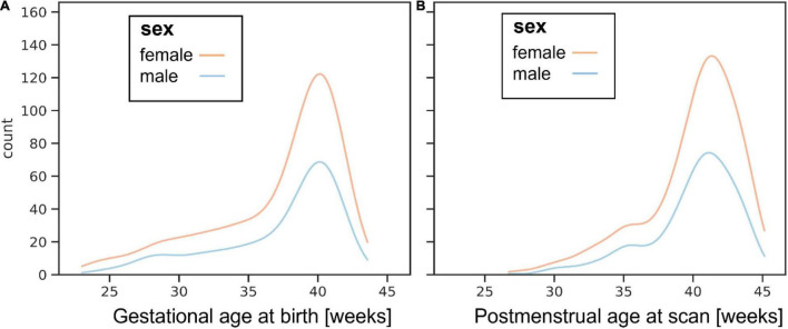
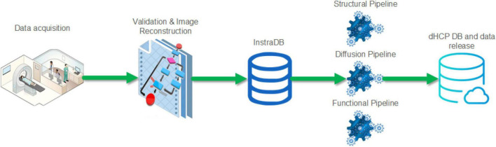
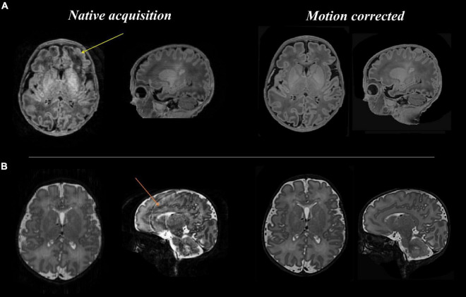
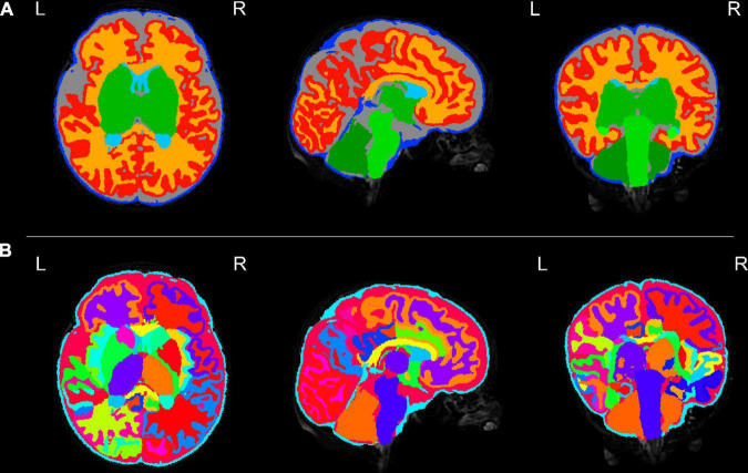
The Developing Human Connectome Project has created a large open science resource which provides researchers with data for investigating typical and atypical brain development across the perinatal period. It has collected 1228 multimodal magnetic resonance imaging (MRI) brain datasets from 1173 fetal and/or neonatal participants, together with collateral demographic, clinical, family, neurocognitive and genomic data from 1173 participants, together with collateral demographic, clinical, family, neurocognitive and genomic data. All subjects were studied in utero and/or soon after birth on a single MRI scanner using specially developed scanning sequences which included novel motion-tolerant imaging methods. Imaging data are complemented by rich demographic, clinical, neurodevelopmental, and genomic information. The project is now releasing a large set of neonatal data; fetal data will be described and released separately. This release includes scans from 783 infants of whom: 583 were healthy infants born at term; as well as preterm infants; and infants at high risk of atypical neurocognitive development. Many infants were imaged more than once to provide longitudinal data, and the total number of datasets being released is 887. We now describe the dHCP image acquisition and processing protocols, summarize the available imaging and collateral data, and provide information on how the data can be accessed.

Keywords: Developing Human Connectome Project; MRI; brain development; connectome; neonatal; perinatal.


Conflict of interest statement

The authors declare that the research was conducted in the absence of any commercial or financial relationships that could be construed as a potential conflict of interest.

Figures

FIGURE 1
FIGURE 1
Histograms showing ages for boys and girls at (A) birth and (B) postnatal MR imaging.
FIGURE 2
FIGURE 2
Schematic of the Developing Human Connectome Project imaging data flow from acquisition to data release.
FIGURE 3
FIGURE 3
Anatomical T1 and T2 weighted images before and after motion correction for one participant. (A: top row) T1 native acquisition (left) with motion artifact visible in the left frontal region in the transverse plane (yellow arrow), which is resolved in the motion corrected images (right) after slice to volume reconstruction. (B: bottom row) T2 native acquisition (left) with motion artifact visible in the sagittal plane (orange arrow), which is resolved in the motion corrected images (right).
FIGURE 4
FIGURE 4
Tissue segmentation and neonatal atlas parcelation for the same infant. Using the automated dHCP structural pipeline, the anatomical images can be segmented into nine tissue classes (A: top row) and parcellated into 87 brain regions (B: bottom row).
FIGURE 5
FIGURE 5
Surface projections using the dHCP structural pipeline for the same infant. (A: top row) 87 region neonatal brain atlas projected onto the pial surface; (B: middle row) Cortical thickness projected onto the inflated cortical surface; and (C: bottom row) Sulcal depth projected onto the inflated cortical surface.
FIGURE 6
FIGURE 6
Resting state functional MRI data from the same infant. (A) An example volume from the fMRI acquisition after image reconstruction and the preprocessing pipeline has been applied; and (B) the auditory and (C) sensorimotor resting state networks. Resting state networks were defined using independent component analysis (ICA) as implemented in FSL MELODIC and have been overlaid onto the native T2 image for ease of visualization.
FIGURE 7
FIGURE 7
Diffusion MRI (dMRI) data from the same infant. Shown are four selected volumes with different b-values and phase encoding directions. Left: input data after MB-SENSE reconstruction. Middle: images after denoising. Right: images after motion and distortion correction and destriping.
FIGURE 8
FIGURE 8
Diffusion MRI metrics in a single subject from the same infant (A) Mean Diffusivity and (B) Color Fractional Anisotropy maps of the Diffusion Tensor Imaging (DTI) model. (C) Tissue Orientation Distribution Function (ODF) of the multi-component analysis in Pietsch et al. (2019). (D) Full brain probabilistic streamline tractography based on the tissue ODF (top image) and based on the mature appearing tissue component (bottom image).
FIGURE 9
FIGURE 9
Probability plot showing age of assessment and combined Bayley III cognitive score for boys and girls.

References

    1. Achenbach T. M., Ruffle T. M. (2000). The child behavior checklist and related forms for assessing behavioral/emotional problems and competencies. Pediatr. Rev. 21 265–271. 10.1542/pir.21-8-265 - DOI - PubMed
    1. Adamson C. L., Alexander B., Ball G., Beare R., Cheong J. L. Y., Spittle A. J., et al. (2020). Parcellation of the neonatal cortex using surface-based Melbourne children’s regional infant brain atlases (M-CRIB-S). Sci. Rep. 10:4359. 10.1038/s41598-020-61326-2 - DOI - PMC - PubMed
    1. Albers C. A., Grieve A. J. (2007). Test review: Bayley, N. (2006). Bayley scales of infant and toddler development-third edition. San Antonio, TX: Harcourt assessment. J. Psychoeduc. Assess. 25 180–190. 10.1177/0734282906297199 - DOI
    1. Alexander-Bloch A., Giedd J. N., Bullmore E. (2013). Imaging structural co-variance between human brain regions. Nat. Rev. Neurosci. 14 322–336. 10.1038/nrn3465 - DOI - PMC - PubMed
    1. Allison C., Baron-Cohen S., Wheelwright S., Charman T., Richler J., Pasco G., et al. (2008). The Q-CHAT (Quantitative CHecklist for Autism in Toddlers): a normally distributed quantitative measure of autistic traits at 18-24 months of age: preliminary report. J. Autism Dev. Disord. 38 1414–1425. 10.1007/s10803-007-0509-7 - DOI - PubMed

LinkOut - more resources