. 2022 Jul 15;25(7):104528.
doi: 10.1016/j.isci.2022.104528. Epub 2022 Jun 3.

VE607 stabilizes SARS-CoV-2 Spike in the "RBD-up" conformation and inhibits viral entry

Affiliations

VE607 stabilizes SARS-CoV-2 Spike in the "RBD-up" conformation and inhibits viral entry

Shilei Ding et al. iScience. .

Abstract

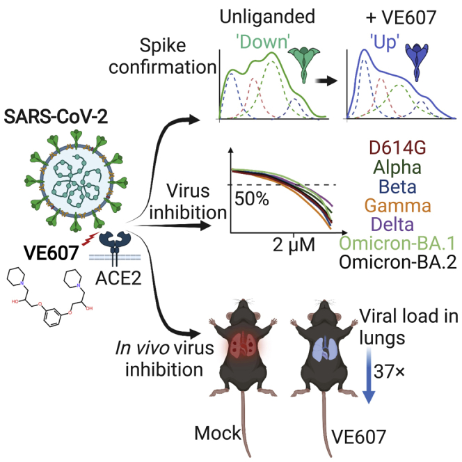
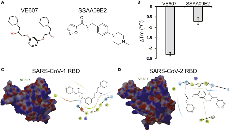
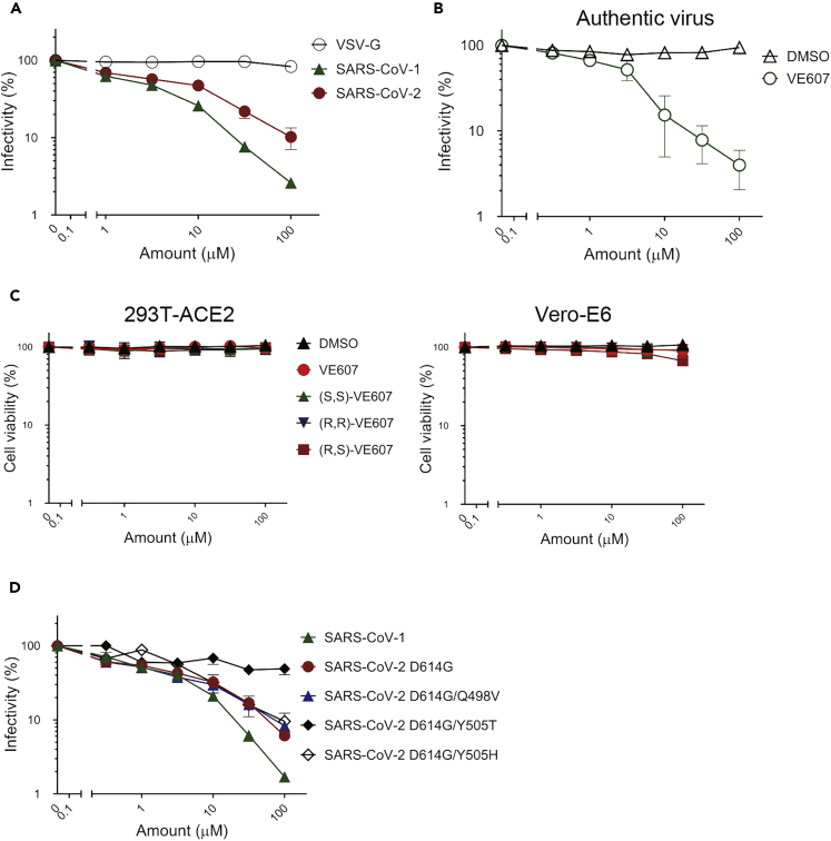
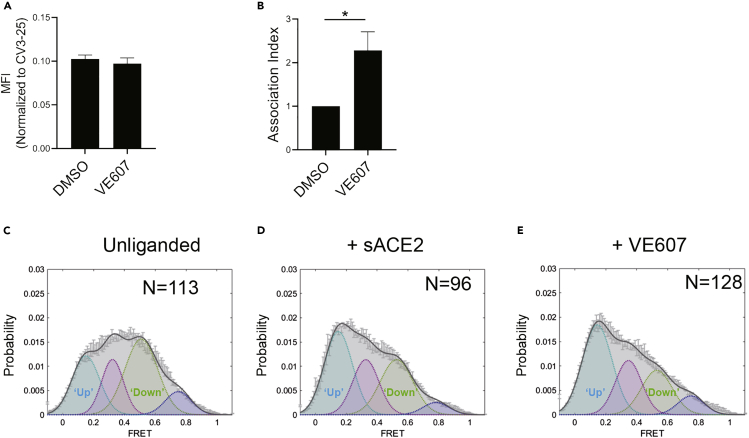
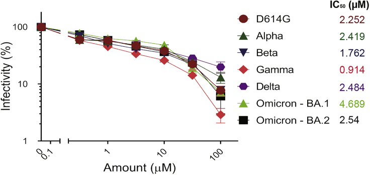
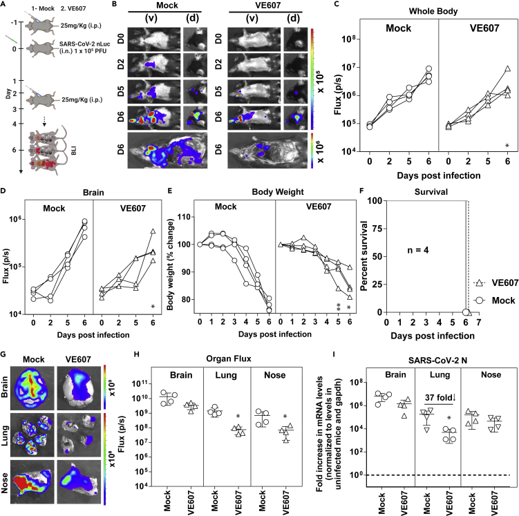
SARS-CoV-2 infection of host cells starts by binding the Spike glycoprotein (S) to the ACE2 receptor. The S-ACE2 interaction is a potential target for therapies against COVID-19 as demonstrated by the development of immunotherapies blocking this interaction. VE607 - a commercially available compound composed of three stereoisomers - was described as an inhibitor of SARS-CoV-1. Here, we show that VE607 broadly inhibits pseudoviral particles bearing the Spike from major VOCs (D614G, Alpha, Beta, Gamma, Delta, Omicron - BA.1, and BA.2) as well as authentic SARS-CoV-2 at low micromolar concentrations. In silico docking, mutational analysis, and smFRET revealed that VE607 binds to the receptor binding domain (RBD)-ACE2 interface and stabilizes RBD in its "up" conformation. Prophylactic treatment with VE607 did not prevent SARS-CoV-2-induced mortality in K18-hACE2 mice, but it did reduce viral replication in the lungs by 37-fold. Thus, VE607 is an interesting lead for drug development for the treatment of SARS-CoV-2 infection.

Keywords: Drugs; Virology.


Conflict of interest statement

The authors declare no competing interests.

Figures

None
Graphical abstract
Figure 1
Figure 1
Potential interactions of SARS-CoV-1 inhibitors with the RBD (A) Chemical structures of VE607 and SSAA09E2. (B) Differential scanning fluorimetry of the SARS-CoV-2 RBD in the presence of SARS-CoV-1 inhibitors, results from two experiments (eight replicates total) are shown. (C) Virtual docking of VE607 to SARS-CoV-1 and (D) SARS-CoV-2 RBD. Left panels, the electrostatic potential is displayed over the molecular surface of the RBD and colored red and blue for negative and positive potential, respectively. Right panels, scheme showing a docking model of VE607 to the RBD. The presumable RBD contact residues are shown as spheres.
Figure 2
Figure 2
VE607 inhibits infection of SARS-CoV-1 and SARS-CoV-2 pseudoviral particles and of authentic SARS-CoV-2 (A) VE607 inhibition of SARS-CoV-1, SARS-CoV-2, or VSV-G (specificity control) pseudovirus. (B) VE607 inhibition of authentic live SARS-CoV-2 virus. (C) VE607 and the three different enantiomers are not toxic on 293T-ACE2 (left) or Vero-E6 (right) cells, as measured by CellTiter-Glo One Solution Assay for the quantitation of ATP presented in live cells. (D) Pseudovirus neutralization of SARS-CoV-2 S mutants predicted by our in silico analysis to modulate the inhibition by VE607. Data represents the average of at least four independent experiments ± SEM.
Figure 3
Figure 3
VE607 stabilizes SARS-CoV-2 S in the “up” conformation (A) VE607 does not compete for sACE2 interaction as measured by flow cytometry. The values represent the median fluorescence intensities (MFI) normalized to binding signals obtained with the conformationally independent CV3-25 Ab. Five experiments are represented as mean ± SEM and statistical significance was tested using unpaired t-test. (B) SARS-CoV-2 Spike stability was measured by radioactive labeling of 293T Spike expressing cells followed by immunoprecipitation of cell lysates and supernatants. At least four experiments are represented as mean ± SEM and statistical significance was tested using unpaired t-test, ∗p < 0.05. (C–E) Single molecule FRET analysis of SARS-CoV-2 S unliganded (C), in presence of sACE2 (D) or VE607 (E).
Figure 4
Figure 4
VE607 inhibits infection of SARS-CoV-2 variants Alpha, Beta, Gamma, Delta, and Omicron pseudovirus particles VE607 inhibits SARS-CoV-2 pseudoviral particles infection of 293T-ACE2 cells. IC50 values are shown next to the Spikes of different VOCs. Data represents the average of at least four independent experiments ± SEM.
Figure 5
Figure 5
VE607 reduces SARS-CoV-2 replication in lungs of K18-hACE2 mice (A) Experimental design to test the efficacy of VE607 in K18-hACE2 mice challenged with SARS-CoV-2-nLuc (WA1, 1 × 105 FFU, i.n.) and treated intraperitoneally (i.p.) from 1 to 4 dpi, (25 mg/kg) with VE607. Vehicle (DMSO)-treated mice were used as controls (Mock). Animals were followed by noninvasive BLI every 2 days as indicated. (B) Representative BLI images of SARS-CoV-2-nLuc-infected mice in ventral (v) and dorsal (d) positions. Scale bars denote radiance (photons/sec/cm2/steradian). (C and D) Temporal quantification of nLuc signal as flux (photons/sec) computed noninvasively. (E) Temporal changes in mouse body weight with initial body weight set to 100% for an experiment shown in A. (F) Kaplan-Meier survival curves of mice (n = 4 per group) statistically compared by log-rank (Mantel-Cox) test for experiment as in A. (G and H) Ex vivo imaging of indicated organs and quantification of nLuc signal as flux (photons/sec) after necropsy. (I) Fold change in SARS-CoV-2 nucleocapsid (N gene) expression in brain, lung, and nose tissues. The data were normalized to Gapdh mRNA in the same sample and that in noninfected mice after necropsy. Viral loads (I) were determined after necropsy at 6 dpi. Each curve in (C–E) and each data point in (C–E) represents an individual mouse. Data in panels are from two independent experiments and n = 2 mice per group. The data in (C–E), (H–I) were analyzed by Mann Whitney nonparametric test. ∗, p < 0.05; ∗∗, p < 0.01; Mean values ± SD are depicted.

Update of

Similar articles

Cited by

References

    1. Adedeji A.O., Severson W., Jonsson C., Singh K., Weiss S.R., Sarafianos S.G. Novel inhibitors of severe acute respiratory syndrome coronavirus entry that act by three distinct mechanisms. J. Virol. 2013;87:8017–8028. doi: 10.1128/jvi.00998-13. - DOI - PMC - PubMed
    1. Anand S.P., Chen Y., Prevost J., Gasser R., Beaudoin-Bussieres G., Abrams C.F., Pazgier M., Finzi A. Interaction of human ACE2 to membrane-bound SARS-CoV-1 and SARS-CoV-2 S glycoproteins. Viruses. 2020;12:1104. - PMC - PubMed
    1. Ball P. The lightning-fast quest for COVID vaccines - and what it means for other diseases. Nature. 2021;589:16–18. doi: 10.1038/d41586-020-03626-1. - DOI - PubMed
    1. Beaudoin-Bussieres G., Laumaea A., Anand S.P., Prevost J., Gasser R., Goyette G., Medjahed H., Perreault J., Tremblay T., Lewin A., et al. Decline of humoral responses against SARS-CoV-2 spike in convalescent individuals. mBio. 2020;11 e02590-20. - PMC - PubMed
    1. Cai Y., Zhang J., Xiao T., Peng H., Sterling S.M., Walsh R.M., Jr., Rawson S., Rits-Volloch S., Chen B. Distinct conformational states of SARS-CoV-2 spike protein. Science. 2020;369:1586–1592. doi: 10.1126/science.abd4251. - DOI - PMC - PubMed

LinkOut - more resources