. 2022 Jun 4;3(2):101444.
doi: 10.1016/j.xpro.2022.101444. eCollection 2022 Jun 17.

Flow cytometry analysis of endothelial cells and subsets of exhausted CD8+ T cells in murine tumor models

Affiliations

Flow cytometry analysis of endothelial cells and subsets of exhausted CD8+ T cells in murine tumor models

Lucas Blanchard et al. STAR Protoc. .

Abstract

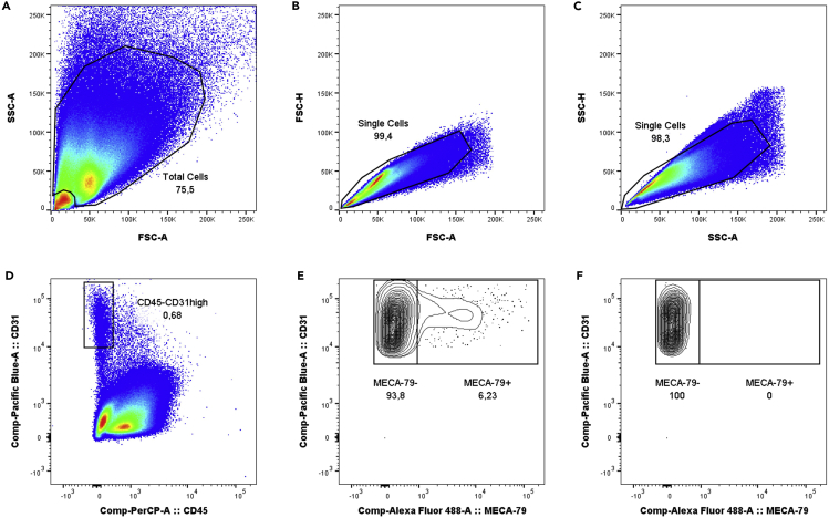
Here, we present a protocol for flow cytometry analysis of endothelial cells (ECs) and CD8+ T cells in murine tumor models, at baseline and after cancer immunotherapy with anti-PD-1/anti-CTLA-4 antibodies. We provide gating strategies for identification of specific cell subsets including ECs from tumor-associated high endothelial venules (TA-HEVs), stem-like, and terminally exhausted CD8+ T cells. This protocol represents a valuable tool for the analysis of rare subsets of tumor ECs and CD8+ T cells with critical roles in antitumor immunity. For complete details on the use and execution of this protocol, please refer to Asrir et al. (2022).

Keywords: Cancer; Cell Biology; Cell isolation; Flow Cytometry/Mass Cytometry; Health Sciences; Immunology; Model Organisms; Single Cell.


Conflict of interest statement

The authors declare no competing interests.

Figures

None
Graphical abstract
Figure 1
Figure 1
Tumor resection (A) Subcutaneous tumor 10 days after inoculation of tumor cells (MCAprog). (B) Visualization of the tumor and the draining lymph node (dLN) following initial dissection.
Figure 2
Figure 2
Tumor digestion (A) Resected tumor (MCAprog) in the collection tube. (B) Minced tumor. (C) Tumor fragments in the digestion tube before enzymatic digestion. (D) Digestion tubes placed on a shaker for enzymatic digestion in a warm room. (E) Tumor cell suspension and remaining fragments after enzymatic digestion. (F) Filtration of the tumor cell suspension and mechanic dissociation of remaining fragments with a syringe plunger in the 50 mL tube overlaid with a cell strainer. (G) Flushing of the cell strainer and syringe plunger. (H) Visualization of the 25 mL of tumor single-cell suspension after entire processing. (I) Tumor-derived cells in the 96-well plate prepared for flow cytometry analysis.
Figure 3
Figure 3
Gating strategy for analysis of tumor endothelial cells (A) Forward and sideward scatter gating to exclude debris. (B) Single cell gating. (C) Single cell gating. (D) CD45-CD31high cell gating to identify total tumor-derived endothelial cells. (E) Identification of MECA-79- tumor-associated endothelial cells (TA-ECs) and MECA-79+ tumor-associated HEV endothelial cells (TA-HECs). (F) FMO MECA-79 control.
Figure 4
Figure 4
Analysis of CD62P and CD62E expression in MECA-79+ TA-HECs (A) Histogram showing CD62P expression in MECA-79+ TA-HECs. (B) Histogram showing CD62E expression in MECA-79+ TA-HECs.
Figure 5
Figure 5
Gating strategy for analysis of tumor-infiltrating CD8+ T cells (A) Single cell gating. (B) Forward and sideward scatter gating to identify lymphoid cells. (C) Live CD45+ cell gating. (D) CD8+ cell gating. (E) CD44+ cell gating. (F) PD-1+ cell gating. (G) Identification of SLAMF6+TIM3- stem-like and SLAMF6-TIM3+ terminally exhausted CD8+ T cells. (H) FMO SLAMF6 control. (I) FMO TIM3 control.
Figure 6
Figure 6
Analysis of TCF-1 and TOX expression in tumor-infiltrating exhausted CD8+ T cells (A) Histograms showing TCF-1 expression in SLAMF6+TIM3- stem-like (SLAMF6+) and SLAMF6-TIM3+ terminally exhausted (TIM3+) CD8+ T cells. (B) Histograms showing TOX expression in SLAMF6+TIM3- stem-like (SLAMF6+) and SLAMF6-TIM3+ terminally exhausted (TIM3+) CD8+ T cells.
Figure 7
Figure 7
Impact of immune checkpoint blockade on tumor-associated endothelial cells (A–D) Representative images of flow cytometry analysis of total tumor-derived endothelial cells (A and C) and MECA-79+ TA-HECs (B and D) in the MCAprog tumor model following treatment with Isotype control (Iso) or combined anti-PD-1 and anti-CTLA-4 antibodies (ICB).
Figure 8
Figure 8
Impact of immune checkpoint blockade on tumor-infiltrating CD8+ T cells (A–H) Representative images of flow cytometry analysis of tumor-infiltrating CD8+ T cells (A and E), PD-1+ CD8+ T cells (B and F), SLAMF6+TIM3- stem-like and SLAMF6-TIM3+ terminally exhausted CD8+ T cells (C and G) and Ki67high SLAMF6+TIM3- stem-like CD8+ T cells (D and H) in the MCAprog tumor model following treatment with Isotype control (Iso) or combined anti-PD-1 and anti-CTLA-4 antibodies (ICB).

References

    1. Asrir A., Tardiveau C., Coudert J., Laffont R., Blanchard L., Bellard E., Veerman K., Bettini S., Lafouresse F., Vina E., et al. Tumor-associated high endothelial venules mediate lymphocyte entry into tumors and predict response to PD-1 plus CTLA-4 combination immunotherapy. Cancer Cell. 2022;40:318–334.e9. doi: 10.1016/j.ccell.2022.01.002. - DOI - PubMed
    1. Blanchard L., Girard J.-P. High endothelial venules (HEVs) in immunity, inflammation and cancer. Angiogenesis. 2021;24:719–753. doi: 10.1007/s10456-021-09792-8. - DOI - PMC - PubMed
    1. Francis D.M., Manspeaker M.P., Schudel A., Sestito L.F., O’Melia M.J., Kissick H.T., Pollack B.P., Waller E.K., Thomas S.N. Blockade of immune checkpoints in lymph nodes through locoregional delivery augments cancer immunotherapy. Sci. Transl. Med. 2020;12:eaay3575. doi: 10.1126/scitranslmed.aay3575. - DOI - PMC - PubMed
    1. Girard J.-P., Moussion C., Forster R. HEVs, lymphatics and homeostatic immune cell trafficking in lymph nodes. Nat. Rev. Immunol. 2012;12:762–773. doi: 10.1038/nri3298. - DOI - PubMed
    1. Khan O., Giles J.R., McDonald S., Manne S., Ngiow S.F., Patel K.P., Werner M.T., Huang A.C., Alexander K.A., Wu J.E., et al. TOX transcriptionally and epigenetically programs CD8+ T cell exhaustion. Nature. 2019;571:211–218. doi: 10.1038/s41586-019-1325-x. - DOI - PMC - PubMed

Publication types

Substances