. 2022 May 23:9:885140.
doi: 10.3389/fcvm.2022.885140. eCollection 2022.

Echocardiography-Guided Percutaneous Patent Ductus Arteriosus Closure: 1-Year Single Center Experience in Indonesia

Affiliations

Echocardiography-Guided Percutaneous Patent Ductus Arteriosus Closure: 1-Year Single Center Experience in Indonesia

Sisca Natalia Siagian et al. Front Cardiovasc Med. .

Abstract

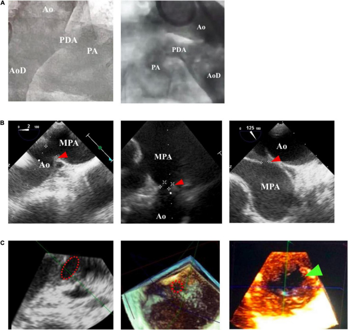
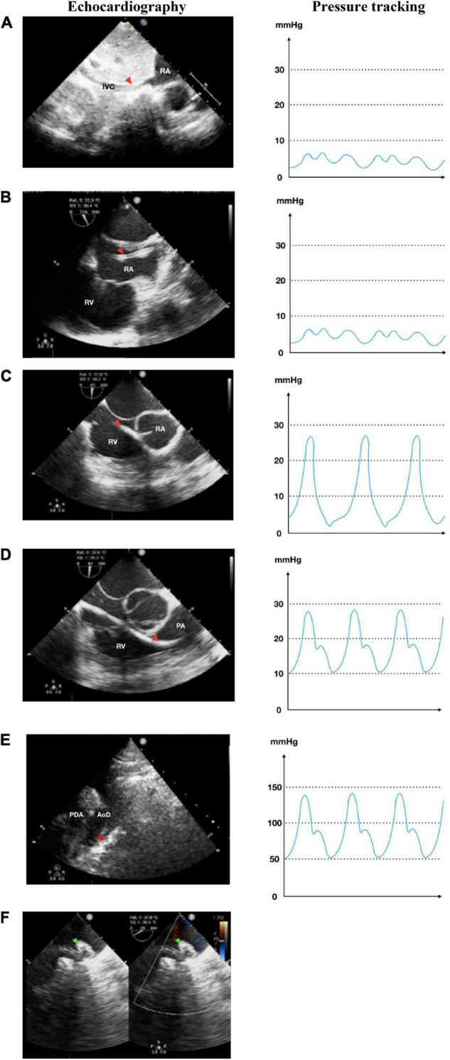
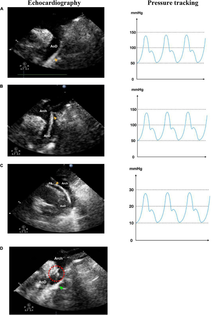
Introduction: Since the first successful percutaneous closure under transesophageal echocardiographic (TEE) guidance, many centers explored transcatheter procedures without fluoroscopy. This single-center study is aimed to show the feasibility and safety of percutaneous patent ductus arteriosus (PDA) closure under echocardiography-only guidance during our 1-year experience.

Methods: Patients with PDA were recruited for percutaneous PDA closure guided by either fluoroscopy or echocardiography-only in National Cardiovascular Center Harapan Kita (ClinicalTrials.gov Identifier: NCT05321849, clinicaltrials.gov/ct2/show/NCT05321849). Patients were evaluated clinically and radiologically using transthoracic echocardiography (TTE) at 6, 24, and 48 h after the procedure. The primary endpoint was the procedural success. Secondary endpoints were the procedural time and the rate of adverse events.

Results: A total of 60 patients underwent transcatheter PDA closure, 30 patients with fluoroscopy and 30 patients with echocardiography guidance. All patients had successful PDA closure. There were only residual shunts, which were disappeared after follow-up in both groups, but one patient with a fluoroscopy-guided procedure had moderate tricuspid regurgitation with suspected thrombus in the tricuspid valve. The procedural time was not significantly different between the fluoroscopy and echocardiography groups.

Keywords: congenital heart disease; echocardiography; fluoroscopy; patent ductus arteriosus; percutaneous.


Conflict of interest statement

The authors declare that the research was conducted in the absence of any commercial or financial relationships that could be construed as a potential conflict of interest.

Figures

FIGURE 1
FIGURE 1
Comparison of patent ductus arteriosus (PDA) size and morphology measurements using fluoroscopy and echocardiography guidance. (A) Fluoroscopy can only provide a 2-dimensional image, whereas (B) transthoracic echocardiography (TTE) and transesophageal echocardiography (TEE) can provide a 3-dimensional structure as shown by the red arrowhead. (C) Cross-sectional PDA morphology measured by using transesophageal echocardiography (TEE), indicated by the dotted red circle. Green arrowhead showed oval-shaped PDA. Ao, aorta; AoD, descending aorta; MPA, main pulmonary artery; PA, pulmonary artery.
FIGURE 2
FIGURE 2
Antegrade transvenous approach of percutaneous patent ductus arteriosus (PDA) closure. (A) Catheter is seen in IVC in transthoracic echocardiography (TTE) subxiphoid view. (B) Catheter is seen in RA in TEE bicaval view (90–110°). (C) Catheter in RV TEE 90° view. (D) Catheter enters PA in TEE 90° view. (E) Catheter crossed from PA towards AoD through PDA in TTE arch view. (F) The device is stowed in place in TEE (40–50°). IVC, inferior vena cava; RA, right atrium; RV, right ventricle; PA, pulmonary artery; Ao, aorta; LA, left atrium; LV, left ventricle; AoD, descending aorta; PDA, patent ductus arteriosus. Red arrowhead shows the catheter head. Green arrowhead indicates the occluder device.
FIGURE 3
FIGURE 3
Retrograde transarterial approach of percutaneous patent ductus arteriosus (PDA) closure. (A) Catheter is seen in AoD in TTE arch view. (B) Catheter is pushed towards the aortic arch in TTE arch view. (C) In parasternal short axis TTE view as high as the great arteries, the catheter is seen in PA through PDA from AoD. (D) In TTE arch view, the device is stowed in place (shown by red dotted circle) after being delivered from the delivery cable. IVC, inferior vena cava; RA, right atrium; RV, right ventricle; PA, pulmonary artery; Ao, aorta; LA, left atrium; LV, left ventricle; AoD, descending aorta; PDA, patent ductus arteriosus. Orange arrowhead shows the catheter head. Green arrowhead indicates the occluder device.

Similar articles

Cited by

References

    1. Baruteau AE, Hascoët S, Baruteau J, Boudjemline Y, Lambert V, Angel CY, et al. Transcatheter closure of patent ductus arteriosus: past, present and future. Arch Cardiovasc Dis. (2014) 107:122–32. 10.1016/j.acvd.2014.01.008 - DOI - PubMed
    1. Hamrick SEG, Hansmann G. Patent ductus arteriosus of the preterm infant. Pediatrics. (2010) 125:1020–30. 10.1542/peds.2009-3506 - DOI - PubMed
    1. Meinel FG, Nance JW, Harris BS, De Cecco CN, Costello P, Schoepf UJ, et al. Radiation risks from cardiovascular imaging tests. Circulation. (2014) 130:442–5. 10.1161/CIRCULATIONAHA.113.005340 - DOI - PubMed
    1. Johnson JN, Sathanandam S, Naik R, Philip R. Echocardiographic guidance for transcatheter patent ductus arteriosus closure in extremely low birth weight infants. Congenit Heart Dis. (2019) 14:74–8. 10.1111/chd.12725 - DOI - PubMed
    1. Baysson H, Nkoumazok B, Barnaoui S, Réhel L, Girodon B, Milani G, et al. Follow-up of children exposed to ionising radiation from cardiac catheterisation: the Coccinelle study. Radiat Prot Dosimetry. (2015) 165:13–6. 10.1093/rpd/ncv039 - DOI - PMC - PubMed

Associated data