Osteochondritis Lesions of the Ischiopubic Area in Young Adolescents
- PMID: 35677753
- PMCID: PMC9170398
- DOI: 10.1155/2022/3573419
Osteochondritis Lesions of the Ischiopubic Area in Young Adolescents
Abstract
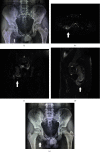
Osteochondritis of the ischiopubic area is a rare disease of children that presents with hip pain and limping. Careful examination and appropriate investigations are essential to establish a definite diagnosis. We report a case series of four children, ages 10-14-year-old, with osteochondritis of the ischiopubic area. Plain X-ray examination showed an area of diffuse irregular calcification of the ischium in two of the children, while in the other two there was an asymmetrical enlargement of the ischiopubic synchondrosis. MRI investigation was the most helpful examination. Bone edema was found in all four children. A calcified mass separated from the host ischium was found in the first two children. The cortex was normal, without irregular destruction. Bone edema of both the ischium and pubic alongside the synchondrosis was found in the following two children, with intact cortices and asymmetrical enlargement. Osteochondritis lesions of the ischium and the ischiopubic area have radiological findings similar to several severe diseases. Bone edema on MRI investigation in children must be properly evaluated. Appropriate radiological examination enabled us to confirm the diagnosis of the osteochondritis and to avoid unnecessary procedures. We want to draw attention to the rare diagnosis of osteochondritis of the ischiopubic area, and the clinical significance, as a cause of hip pain and limping in children.
Copyright © 2022 Nikolaos Laliotis et al.
Conflict of interest statement
The authors declare that they have no conflict of interest.
Figures


References
-
- Achar S., Yamanaka J. Apophysitis and osteochondrosis: common causes of pain in growing bones. American Family Physician . 2019;99(10):610–618. - PubMed
-
- Vassalou E. E., Karantanas A. H., Raissaki M. Imaging of traumatic injuries of the paediatric pelvis and hip. Hellenic Journal οf Radiology . 2018;3(3):20–40.
-
- Schneider K. N., Lampe L. P., Gosheger G., et al. Invasive diagnostic and therapeutic measures are unnecessary in patients with symptomatic van Neck-Odelberg disease (ischiopubic synchondrosis): a retrospective single-center study of 21 patients with median follow-up of 5 years. Acta Orthopaedica . 2021;92(3) - PMC - PubMed
Publication types
LinkOut - more resources
Full Text Sources

