. 2022 May 23:16:890827.
doi: 10.3389/fncel.2022.890827. eCollection 2022.

Early Developmental PMCA2b Expression Protects From Ketamine-Induced Apoptosis and GABA Impairments in Differentiating Hippocampal Progenitor Cells

Affiliations

Early Developmental PMCA2b Expression Protects From Ketamine-Induced Apoptosis and GABA Impairments in Differentiating Hippocampal Progenitor Cells

Malwina Lisek et al. Front Cell Neurosci. .

Abstract

PMCA2 is not expressed until the late embryonic state when the control of subtle Ca2+ fluxes becomes important for neuronal specialization. During this period, immature neurons are especially vulnerable to degenerative insults induced by the N-methyl-D-aspartate (NMDA) receptor blocker, ketamine. As H19-7 hippocampal progenitor cells isolated from E17 do not express the PMCA2 isoform, they constitute a valuable model for studying its role in neuronal development. In this study, we demonstrated that heterologous expression of PMCA2b enhanced the differentiation of H19-7 cells and protected from ketamine-induced death. PMCA2b did not affect resting [Ca2+]c in the presence or absence of ketamine and had no effect on the rate of Ca2+ clearance following membrane depolarization in the presence of the drug. The upregulation of endogenous PMCA1 demonstrated in response to PMCA2b expression as well as ketamine-induced PMCA4 depletion were indifferent to the rate of Ca2+ clearance in the presence of ketamine. Yet, co-expression of PMCA4b and PMCA2b was able to partially restore Ca2+ extrusion diminished by ketamine. The profiling of NMDA receptor expression showed upregulation of the NMDAR1 subunit in PMCA2b-expressing cells and increased co-immunoprecipitation of both proteins following ketamine treatment. Further microarray screening demonstrated a significant influence of PMCA2b on GABA signaling in differentiating progenitor cells, manifested by the unique regulation of several genes key to the GABAergic transmission. The overall activity of glutamate decarboxylase remained unchanged, but Ca2+-induced GABA release was inhibited in the presence of ketamine. Interestingly, PMCA2b expression was able to reverse this effect. The mechanism of GABA secretion normalization in the presence of ketamine may involve PMCA2b-mediated inhibition of GABA transaminase, thus shifting GABA utilization from energetic purposes to neurosecretion. In this study, we show for the first time that developmentally controlled PMCA expression may dictate the pattern of differentiation of hippocampal progenitor cells. Moreover, the appearance of PMCA2 early in development has long-standing consequences for GABA metabolism with yet an unpredictable influence on GABAergic neurotransmission during later stages of brain maturation. In contrast, the presence of PMCA2b seems to be protective for differentiating progenitor cells from ketamine-induced apoptotic death.

Keywords: GABA metabolism; calcium; hippocampal progenitor cells; ketamine; neuronal differentiation; plasma membrane Ca2+-ATPase (PMCA).


Conflict of interest statement

The authors declare that the research was conducted in the absence of any commercial or financial relationships that could be construed as a potential conflict of interest.

Figures
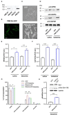
Figure 1
Figure 1
PMCA2b expression enhances H19-7 cell differentiation and restricts ketamine-mediated apoptosis. (A) The presence of PMCA isoforms determined using the Western blot. (B) The expression of PMCA2b-GFP and the targeting of the fusion protein to the plasma membrane visualized using immunocytochemistry. Scale bar 20 μm. (C) The effect of PMCA2b expression on cellular morphology in the absence of a differentiating agent. Scale bar 50 μm. (D) The protein level of sdifferentiation markers, NF68 and GAP43, determined using the Western blot. For (A–D), the representative images are presented. (E) Quantification of NF68 and (F) GAP43 band intensity following normalization to endogenous Gapdh level, n = 4. AU, arbitrary units. (G) Flow cytometry analysis of differentiated cell viability in the presence or absence of ketamine. (H) Quantification of Bad phosphorylation at Ser-136. Total Bad and p-Bad Ser-136 protein level determined by the Western blot in the presence of ketamine was normalized to Gapdh, and the results are presented as phosphorylation index (pBad/Bad), n = 3. Representative blots are shown. *P < 0.05, **P < 0.01, ***P < 0.001.
Figure 2
Figure 2
The effect of PMCA2b expression on KCl-induced Ca2+ transients in the presence or absence of ketamine. (A) Representative traces of Fluo-4 fluorescence intensity (F/F0) changes in response to 30 mM KCl pulse measured in single cells using Leica DMi8 inverted microscope. (B) Quantification of a resting Ca2+ in cytosol ([Ca2+]c), n = 17. (C) Halftime of Ca2+ signal decay, n = 17. For (B,C), the data are plotted from max to min with the line centered at the mean. *P < 0.05, ***P < 0.001.
Figure 3
Figure 3
The contribution of PMCA isoforms to the rate of Ca2+ clearance following 30 mM KCl treatment in the presence or absence of ketamine. (A) The expression of Atp2b1 in GFP- or PMCA2b-GFP expressing cells was qualified using real-time PCR. The raw data were normalized to endogenous Gapdh expression and were calculated based on the 2−ΔΔCt method to obtain relative fold change. The expression level in GFP control treated with saline was taken as 1, n = 4. (B) PMCA1 protein level was determined with the Western blot. GAPDH was used as a loading control. The representative images are shown. (C) Quantification of PMCA1 protein level following normalization to endogenous Gapdh level, n = 3. AU, arbitrary units. (D) The efficiency of Atp2b1 silencing with siRNA evaluated using real-time PCR and confirmed at the protein level with the Western blot. The reduction in mRNA level was quantified using the 2−ΔΔCt method, and the expression in scrambled-treated cells was taken as 1, n = 3. (E) Halftime of signal decay (t1/2) of individual tracings recorded from scrambled or PMCA1 siRNA-treated cells, presented as interleaved box graph centered at the mean. (F) The expression of Atp2b4 in GFP- or PMCA2b-GFP expressing cells, n = 3. (G) Immunoreactivity of PMCA4 determined with the Western blot. (H) Quantification of PMCA4 protein level following normalization to endogenous Gapdh level, n = 3. AU, arbitrary units. (I) Over-expression of PMCA4b-mCherry protein and the targeting of the fusion protein to the plasma membrane visualized by immunocytochemistry. Scale bar 20 μm. (J) Halftime of signal decay (t1/2) of individual tracings recorded from mCherry- or PMCA4b-mCherry-positive cells, presented as interleaved box graph centered at the mean. Here, the mock-transfected control was cells double transfected with GFP and mCherry. *P < 0.05, **P < 0.01, ***P < 0.001.
Figure 4
Figure 4
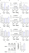
Ketamine enhances PMCA2b interaction with NMDAR1. (A) The expression of Grin1 encoding NMDAR1 subunit evaluated with real-time PCR and calculated using the 2−ΔΔCt method. The expression level in GFP control was taken as 1, n = 4. (B) NMDAR1 protein level evaluated with the Western blot. GAPDH was taken as a loading control. The representative blots are presented. (C) Quantification of NMDAR1 protein level following normalization to endogenous Gapdh level, n = 3. AU, arbitrary units. (D) The mRNA and (E) protein levels of NMDAR2a. (F) Quantification of NMDAR2a protein level following normalization to endogenous Gapdh level, n = 3. (G) The mRNA and (H) protein levels of NMDAR2b. (I) Quantification of NMDAR2b protein level following normalization to endogenous Gapdh level, n = 3. (J) Co-immunoprecipitation of NMDAR1 and PMCA2b. Lysates from differentiating H19-7 cells were used to measure NMDAR1 protein level (input) in saline- and ketamine-treated conditions. Negative controls also included sepharose-linked secondary antibodies (IgG) and sepharose beads only. Representative blots are presented. (K) Quantitative densitometric analysis of band intensity in PMCA2b-expressing cells. The results are presented as arbitrary units defined as the optical density per mg protein, n = 3. *P < 0.05, **P < 0.01.
Figure 5
Figure 5
Microarray screening of fundamental genes involved in glutamate and GABA signaling. The SABiosciences rat microarray (PARN-152Z) was used to evaluate changes in the expression of genes involved in GABA and glutamate signaling. [(A–D), left panel] Volcano plot analysis of expression changes calculated using company-provided software. [(A–D), right panel] Genes whose expression was statistically significant (P < 0.05).
Figure 6
Figure 6
The effect of ketamine on GABA metabolism in the presence or absence of PMCA2b. (A) The activity of GAD assayed based on the fluorometric measurement of GABA and ninhydrin condensation product. n = 4. (B) Quantification of Ca2+-dependent GABA release over 5 min stimulation with 30 mM KCl done with GABA Elisa kit. n = 3. (C) The activity of GABA transaminase (GABA-T) expressed as NADH formation measured at 340 nm. n = 4. *P < 0.05, **P < 0.01, ***P < 0.001.

Similar articles

References

    1. Abekawa T., Ito K., Nakagawa S., Koyama T. (2007). Prenatal EXPOSURE to an NMDA Receptor antagonist, MK-801 reduces density of parvalbumin-immunoreactive GABAergic neurons in the medial prefrontal cortex and enhances phencyclidine-induced hyperlocomotion but not behavioral sensitization to methamphetamine in postpubertal rats. Psychopharmacology 192, 303–316. 10.1007/s00213-007-0729-8 - DOI - PubMed
    1. Afroze T., Husain M. (2001). Cell CYCLE dependent regulation of intracellular calcium concentration in vascular smooth muscle cells: a potential target for drug therapy. Curr. Drug Targets Cardiovasc. Haematol. Disord. 1, 23–40. 10.2174/1568006013338060 - DOI - PubMed
    1. Aligny C., Roux C., Dourmap N., Ramdani Y., Do,-REGO J. C., Jégou S., et al. . (2014). Ketamine alters cortical integration of GABAergic interneurons and induces long-term sex-dependent impairments in transgenic Gad67-GFP mice. Cell Death Dis. 5, e1311. 10.1038/cddis.2014.275 - DOI - PMC - PubMed
    1. Augustine G. J., Santamaria F., Tanaka K. (2003). Local calcium signaling in neurons. Neuron 40, 331–346. 10.1016/S0896-6273(03)00639-1 - DOI - PubMed
    1. Behrens M. M., Ali S. S., Dao D. N., Lucero J., Shekhtman G., Quick K. L., et al. . (2007). Ketamine-induced loss of phenotype of fast-spiking interneurons is mediated by NADPH-oxidase. Science 318, 1645–1647. 10.1126/science.1148045 - DOI - PubMed