Case Reports
. 2022 Jun 3:16:107-111.
doi: 10.1016/j.artd.2022.05.004. eCollection 2022 Aug.

Save the Greater Trochanter: A Novel Modification to the Extended Trochanteric Osteotomy

Affiliations
Case Reports

Save the Greater Trochanter: A Novel Modification to the Extended Trochanteric Osteotomy

Eric B Smith. Arthroplast Today. .

Abstract

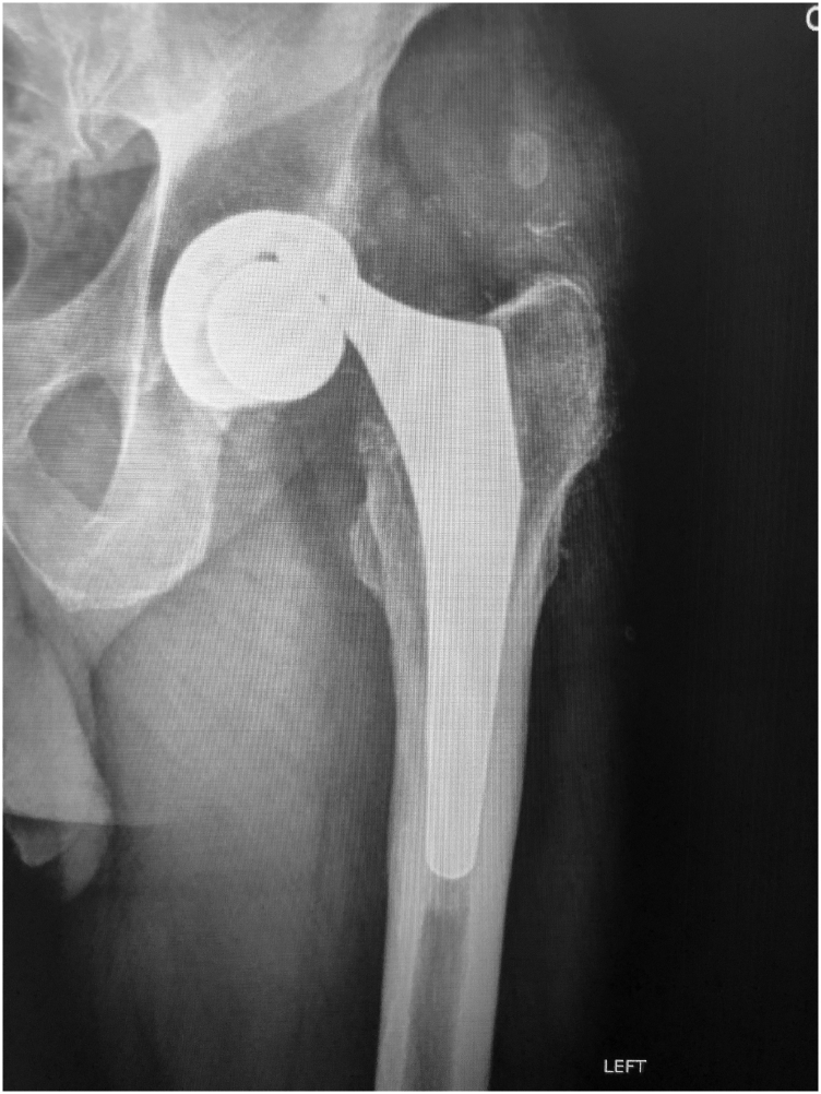
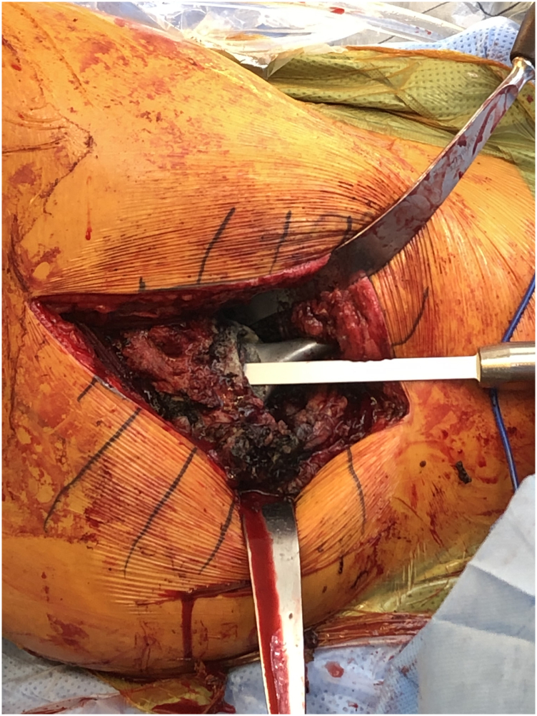
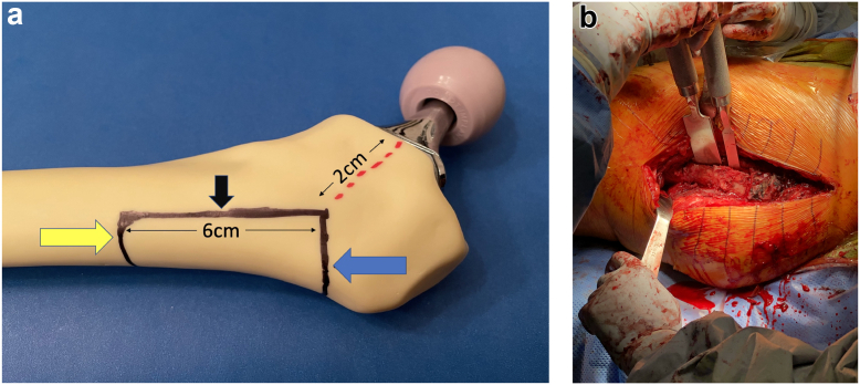
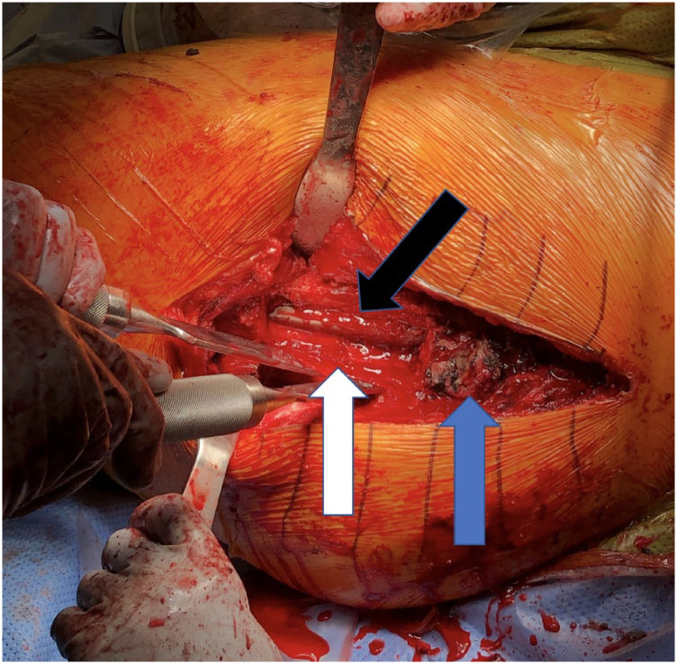
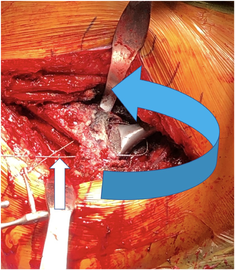
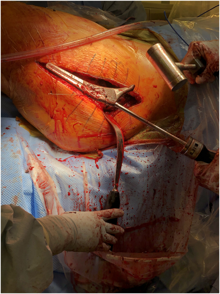
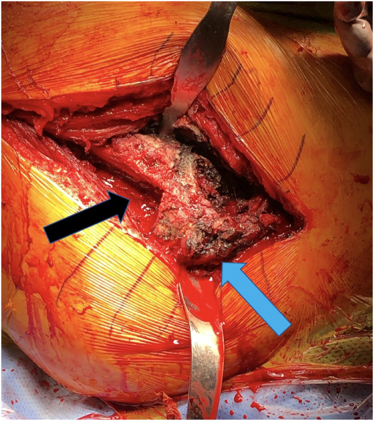
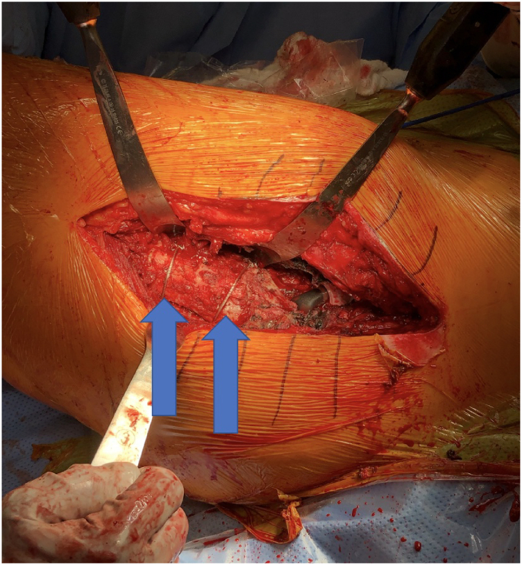
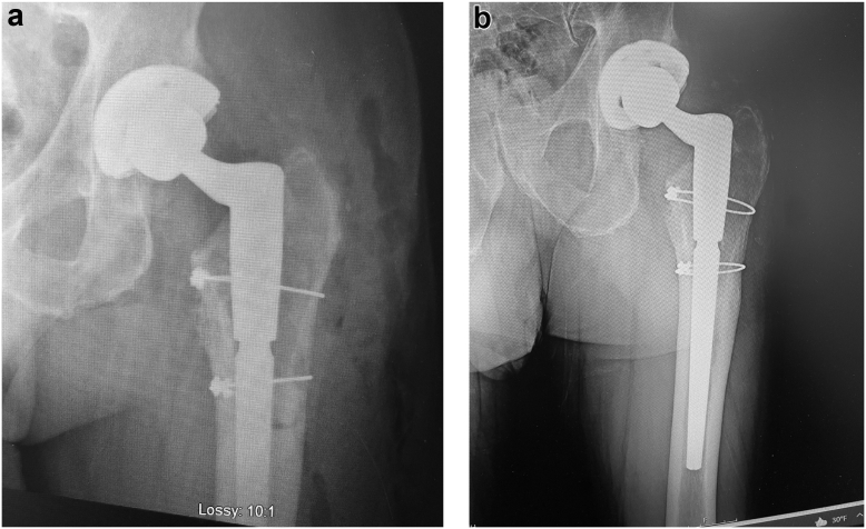
The extended trochanteric osteotomy is the workhorse for removal of well-fixed femoral stems during total hip revision arthroplasty. Despite its reliable performance in exposing the implants for removal and accessing the femoral canal, significant complications can occur. Though these complications are rare, trochanteric nonunion, trochanteric escape, and femoral implant subsidence can have a significant negative impact on gait mechanics and patient outcome. If access to the canal was still possible and the greater trochanter could remain in place, these complications could be minimized or possibly even eliminated. This paper describes a novel technique using a lateral cortical window just distal to the greater trochanter that allows removal of a well-fixed stem and leaves the greater trochanter intact.

Keywords: Extended trochanteric osteotomy; Femoral stem removal; Surgical technique; Total hip revision.


Figures

Figure 1
Figure 1
Preoperative AP radiograph with catastrophic failure of trunnion with complete dissociation of the cobalt chromium femoral head. The damaged stem required removal. Though minor radiographic signs are present that may indicate a loose stem, it was found to be well fixed at the time of surgery. Before possibly fracturing the proximal femur, the modified ETO technique was performed.
Figure 2
Figure 2
Intraoperative photograph with hip dislocated during a direct lateral approach. The patient is in supine position with his head to the right and feet to the left. After passing a small burr circumferentially around the proximal stem, a flexible osteotome was passed distally around the stem extending at least 2 cm without perforating the cortex. Attempts were made to distract the stem without success.
Figure 3
Figure 3
A saw bones replica of the proximal femur (a). The proximal border (blue arrow) of the osteotomy is just distal to vastus ridge and leaving at least 2 cm of proximal bone. The distal border (yellow arrow) is measured 6 cm distally from the proximal border (based on the porous coating of this stem). The anterior border (black arrow) connects the proximal and distal borders along the lateral edge of the stem. Intraoperative photograph (b) after the anterior cut was made with the oscillating saw and the proximal and distal cuts were made with a pencil tip burr. The posterior cut was made passing the saw through the anterior cut to score the inner cortex. Osteotomes were used to lever out the fragment.
Figure 4
Figure 4
The osteotomized fragment (white arrow) is retracted posteriorly. The greater trochanter (blue arrow) is completely intact. The implant (black arrow) and distal aspect of the porous coating is visualized within the femoral canal.
Figure 5
Figure 5
The Gigli saw (white arrow) is passed through the osteotomy site up and over the neck of the stem (blue arrow). It may be necessary to use a burr or osteotome through the osteotomy site directed proximally if there is any obstruction to passing the Gigli saw. The saw is then passed back and forth moving distally around stem until all bony ingrowth/outgrowth is released.
Figure 6
Figure 6
Stem removed with no appreciable bone attachment.
Figure 7
Figure 7
Proximal femur after stem removal. The osteotomy site (black arrow) and greater trochanter (blue arrow) are intact and without damage.
Figure 8
Figure 8
Proximal femur after osteotomy repair with 2 metallic cables (blue arrows).
Figure 9
Figure 9
Immediate postoperative AP radiograph (a) with modular cone/conical cylindrical stem. Osteotomy site is visible with 2 cerclage cables holding in anatomic position. One-year postoperative AP radiograph (b) showing complete anatomic healing of the osteotomy fragment and no subsidence of the stem.

Similar articles

Cited by

References

    1. Lerch M., von Lewinski G., Windhagen H., Thorey F. Revision of total hip arthroplasty: clinical outcome of extended trochanteric osteotomy and intraoperative femoral fracture. Technol Health Care. 2008;16:293–300. - PubMed
    1. Mardones R., Gonzalez C., Cabanela M.E., Trousdale R.T., Berry D.J. Extended femoral osteotomy for revision of hip arthroplasty: results and complications. J Arthroplasty. 2005;20:79–83. - PubMed
    1. Park Y.S., Moon Y.W., Lim S.J. Revision total hip arthroplasty using a fluted and tapered modular distal fixation stem with and without extended trochanteric osteotomy. J Arthroplasty. 2007;22:993–999. - PubMed
    1. Wronka K.S., Gerard-Wilson M., Peel E., Rolfson O., Cnudde P.H.J. Extended trochanteric osteotomy: improving the access and reducing the risk in revision THA. EFORT Open Rev. 2020;5:104–112. - PMC - PubMed
    1. MacDonald S.J., Cole C., Guerin J., Rorabeck C.H., Bourne R.B., McCalden R.W. Extended trochanteric osteotomy via the direct lateral approach in revision hip arthroplasty. Clin Orthop Relat Res. 2003;417:210–216. - PubMed

Publication types

LinkOut - more resources