Review
. 2022 Aug;9(23):e2201160.
doi: 10.1002/advs.202201160. Epub 2022 Jun 9.

Förster Resonance Energy Transfer in Luminescent Solar Concentrators

Affiliations
Review

Förster Resonance Energy Transfer in Luminescent Solar Concentrators

Bolong Zhang et al. Adv Sci (Weinh). 2022 Aug.

Abstract

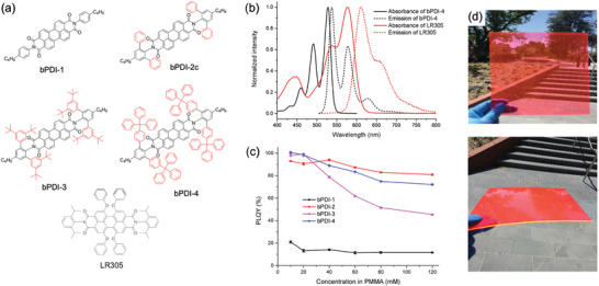
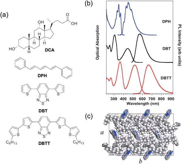
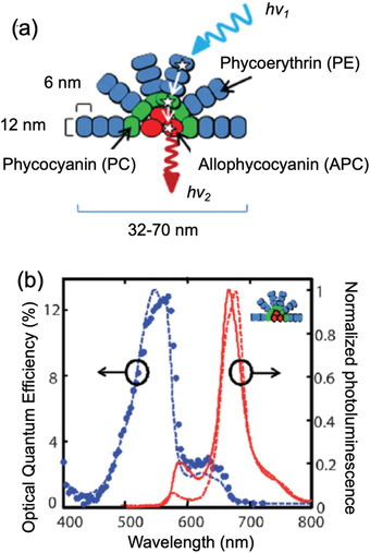
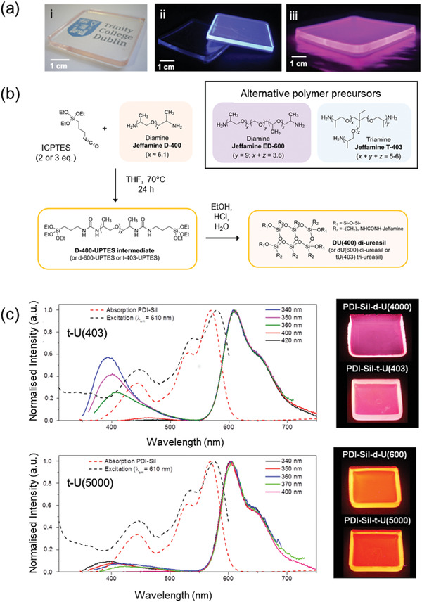
Luminescent solar concentrators (LSCs) are an emerging technology to collect and channel light from a large absorption area into a smaller one. They are a complementary technology for traditional solar photovoltaics (PV), particularly suitable for application in urban or indoor environments where their custom colors and form factors, and performance under diffuse light conditions may be advantageous. Förster resonance energy transfer (FRET) has emerged as a valuable approach to overcome some of the intrinsic limitations of conventional single lumophore LSCs, such as reabsorption or reduced quantum efficiency. This review outlines the potential of FRET to boost LSC performance, using highlights from the literature to illustrate the key criteria that must be considered when designing an FRET-LSC, including both the photophysical requirements of the FRET lumophores and their interaction with the host material. Based on these criteria, a list of design guidelines intended to aid researchers when they approach the design of a new FRET-LSC system is presented. By highlighting the unanswered questions in this field, the authors aim to demonstrate the potential of FRET-LSCs for both conventional solar-harvesting and emerging LSC-inspired technologies and hope to encourage participation from a diverse researcher base to address this exciting challenge.

Keywords: Förster resonance energy transfer; light harvesting; luminescent solar concentrator; lumophore; solar energy.


Conflict of interest statement

The authors declare no conflict of interest.

Figures

Figure 1
Figure 1
Working mechanism and prototype of an LSC application. a) A typical LSC consists of a lightguide slab that is doped or coated with lumophores. The lumophores absorb incident light and re‐emit it as photoluminescence. The emitted photons are transported by total internal reflectance to the slab edges where they can be collected by attached photovolatic cells. The LSC efficiency is decreased by losses such as transmittance (non‐absorption of incident light), reflectance of incident or emitted light from internal/external surfaces, and reabsorption of emitted photons by neighboring lumophores in the emission path. b) Photograph of a prototype LSC smart‐window (≈30 × 42 cm2) charging a cell phone.
Figure 2
Figure 2
Mechanism of FRET. a) Simplified Jablonski diagram of a typical FRET process following light absorption by the donor. b) FRET helps overcome reabsorption losses, which are common in single lumophore systems exhibiting a small Stokes shift (green—upper). The addition of an emitter (pink) helps reduce reabsorption by increasing the effective Stokes shift. Just a small emitter:donor ratio is required to fully quench the donor's photoluminescence, and the emitter becomes the primary emissive species. c) Schematic representation of FRET between donor and emitter molecules in a lightguide host (black rectangle). Donor molecules absorb the incident light and energy may pass between several donor molecules via energy migration prior to FRET to the emitter species.
Figure 3
Figure 3
Energy transfer cascades in a tri‐lumophore intermolecular FRET system based on the two conjugated polymer donors (P1, P2) and a molecular emitter (Red 305). a) Proposed energy transfer mechanism. b) Chemical structures and c) absorption and photoluminescence spectra of the donor/emitter species. Adapted with permission.[ 34 ] Copyright 2016, Wiley‐VCH.
Figure 4
Figure 4
The addition of bulky substituents can inhibit ACQ in perylene diimides. a) Chemical structure, b) absorption and emission spectra, and c) concentration‐dependent PLQY of different perylene diimide structures. d) Photograph of large‐area LSCs (20 × 20 × 0.1 cm3) based on the bPDI‐3/LR305 FRET pair. a‐c) Reproduced with permission.[ a]Copyright 2017, American Chemical Society. d) Reproduced with permission.[16c] Copyright 2019, American Chemical Society.
Figure 5
Figure 5
AIEgens can be deployed effectively in FRET‐LSCs. a,b) Chemical structures of reported AIEgens used in FRET‐LSCs. c) Photographs of donor (p‐O‐TPE), donor–emitter (p‐O‐TPE‐PDI‐Sil), and emitter (PDI‐Sil) lumophores in a ureasil (du(600)) lightguide under natural (top) and UV (bottom) light. Adapted with permission.[ 48 ] Copyright 2019, American Chemical Society.
Figure 6
Figure 6
A tandem thin‐film LSC based on FRET layers. a) Architecture of the tandem LSCs, where the top layer absorbs incident light at short wavelengths and the bottom layer absorbs longer wavelengths. b) The chemical structures of rubrene (donor), DCJTB (emitter), and Pt(TPBP) (emitter). c) The normalized absorption and emission spectra of the FRET‐LSC and phosphorescent LSC, respectively. Adapted with permission.[ 16b ] Copyright 2008, American Association for the Advancement of Science. The reabsorption factor, S, was defined as the ratio of the absorbance at the absorption and emission maxima of the lumophore system.
Figure 7
Figure 7
Self‐assembly can be used to localize FRET pairs. a) Chemical structure of the host material DCA and the three lumophores used in the FRET cascade. b) The absorption and emission spectra of the three lumophores. c) Structure of DPH–DCA host–guest system. Adapted with permission.[ 52 ] Copyright 2013, Royal Society of Chemistry.
Figure 8
Figure 8
Polymer and dendrimer‐based intramolecular FRET systems. a) Chemical structures of the donor, emitter, and intramolecular FRET trimers reported by Webb et al. The inset figure shows the absorption spectra of the emitters overlap well with the PL spectrum of the donor. Adapted with permission.[ 57 ] Copyright 2016, Royal Society of Chemistry. b) Chemical structures of the copolymers, and the donor and emitter monomers of the FRET system reported by Davis et al., and the corresponding absorption and emission spectra of the donor and emitter monomers. Adapted with permission.[ 56 ] Copyright2016, Royal Society of Chemistry. c) Chemical structures of the BODIPY monomers and corresponding FRET dendrimer reported by Bozdemir et al. Adapted with permission.[ 60 ] Copyright 2011, Wiley‐VCH.
Figure 9
Figure 9
An FRET dendrimer for LSCs. a) Chemical structures of the three FRET dendrimers based‐on a BODIPY core with three oligofluorene side‐chains. b) Absorption and emission spectra of the FRET dendrimers. c) Chemical structures of hypothetical FRET dendrimers structures predicted to show improved LSC performance through Monte‐Carlo simulations. Adapted with permission.[ 62 ] Copyright 2017, Royal Society of Chemistry.
Figure 10
Figure 10
A FRET‐LSC based on a natural light‐harvesting antenna system. a) The structure of the phycobilisome consists of three lumophores self‐assembled in an FRET cascade. b) Absorption and PL spectra of the phycobilisome. Adapted with permission.[ 63 ] Copyright 2009, Wiley‐VCH.
Figure 11
Figure 11
An FRET‐based liquid LSC using surface‐functionalized QD lumophores. a) Schematic representation of the liquid LSC architecture (top) and the FRET process between Qdots 545 and the surface‐anchored AFDye. b) Reaction scheme for covalent grafting of the AFDye to the coating of the QDs. Adapted with permission.[ 16d ] Copyright 2017, Elsevier.
Figure 12
Figure 12
Photoactive lightguides based on ureasils can participate in FRET processes in LSCs. a) Photographs of representative ureasil samples under i) visible light and UV illumination for ii) pure and single lumophore doped ureasils and iii) a two‐lumophore FRET sample. Adapted with permission. Adapted with permission.[ 73b ] Copyright 2017, Royal Society of Chemistry. b) Representative synthetic route to a (di)‐ureasil hybrid. The structure can be varied by changing the polymer precursor. Adapted with permission.[ 71 ] Copyright 2020, SPIE. c) Absorption/excitation and emission spectra of PDI‐Sil doped tri‐ureasils. The efficiency of energy transfer from the ureasil host (≈370–500 nm) to the PDI‐Sil emitter (≈550–750 nm) can be controlled by varying the molecular weight of the polymer backbone (e.g., from 403 to 5000 g mol−1), as demonstrated by the degree of quenching of the ureasil emission and the observed total sample emission (see photographs on right). Adapted with permission.[ 74d] Copyright 2016, Royal Society of Chemistry.
Figure 13
Figure 13
The intensity of absorption and emission depends on lumophore alignment. a) In planar organic lumophores, the transition dipole moment (TDM) is located along the long axis of the molecule. Absorption is favored when the electric field vector of the incident photon is parallel to the TDM (i.e., perpendicular to the direction of travel), and unflavored in the orthogonal axis. Similarly, photoluminescence is enhanced under the same conditions. Adapted with permission.[ 50 ] Copyright 2019, American Chemical Society. b) An example of a dichroic molecule, Rhodamine 6G, showing the orientation of the transition dipole moment. c) Lumophore alignment in an LSC will affect the absorption and emission properties. In the vertically aligned LSC, the TDM of the dichroic lumophore is perpendicular to the substrate surface, leading to reduced absorption. In the horizontal alignment, the TDM is parallel to the substrate surface and, quite often, two opposite edges of the LSC, which can lead to reabsorption losses.
Figure 14
Figure 14
Poor light absorption in vertically‐aligned LSCs can be overcome using a non‐dichroic donor with a dichroic emitter. a) Schematic diagram of the OEP (donor)/R800 (emitter) FRET pair vertically aligned by the liquid crystal matrix, and b) the decoherence of the degenerated transition dipole pair in OEP. One dipole absorbs the incident light and the second transfers energy to the emitter via FRET. Adapted with permission.[ 81 ] Copyright 2013, American Chemical Society.
Figure 15
Figure 15
Tunable alignment of molecular lumophores in liquid crystal matrices. a) A FRET‐LSC based on rod‐shaped emitters aligned vertically the liquid crystal matrix, while the spherical donors remained in random orientation to absorb the incident light. b) Chemical structures and c) molecular shapes of the emitter and donors. Adapted with permission.[ 50 ] Copyright 2019, American Chemical Society. d) A switchable LSC in which the lumophore alignment can be switched between i) absorbing and ii) transparent modes upon application of an electric field. iii) Chemical structure of the donor–emitter‐donor intramolecular FRET triad used as the lumophore in this LSC. Adapted with permission.[ 78 ] Copyright 2014, American Chemical Society.

References

    1. Renewable Capacity Highlights, IRENA 2020.
    1. Meinardi F., Bruni F., Brovelli S., Nat. Rev. Mater. 2017, 2, 17072.
    1. Rafiee M., Chandra S., Ahmed H., McCormack S. J., Opt. Mater. 2019, 91, 212.
    1. Li Y., Zhang X., Zhang Y., Dong R., Luscombe C. K., J. Polym. Sci., Part A: Polym. Chem. 2019, 57, 201.
    1. Weber W. H., Lambe J., Appl. Opt. 1976, 15, 2299. - PubMed

Publication types

MeSH terms

LinkOut - more resources