Review
. 2022 Jun 1;28(3):750-780.
doi: 10.1212/CON.0000000000001124.

Vascular Cognitive Impairment and Dementia

Review

Vascular Cognitive Impairment and Dementia

Ellen Chang Wong et al. Continuum (Minneap Minn). .

Abstract

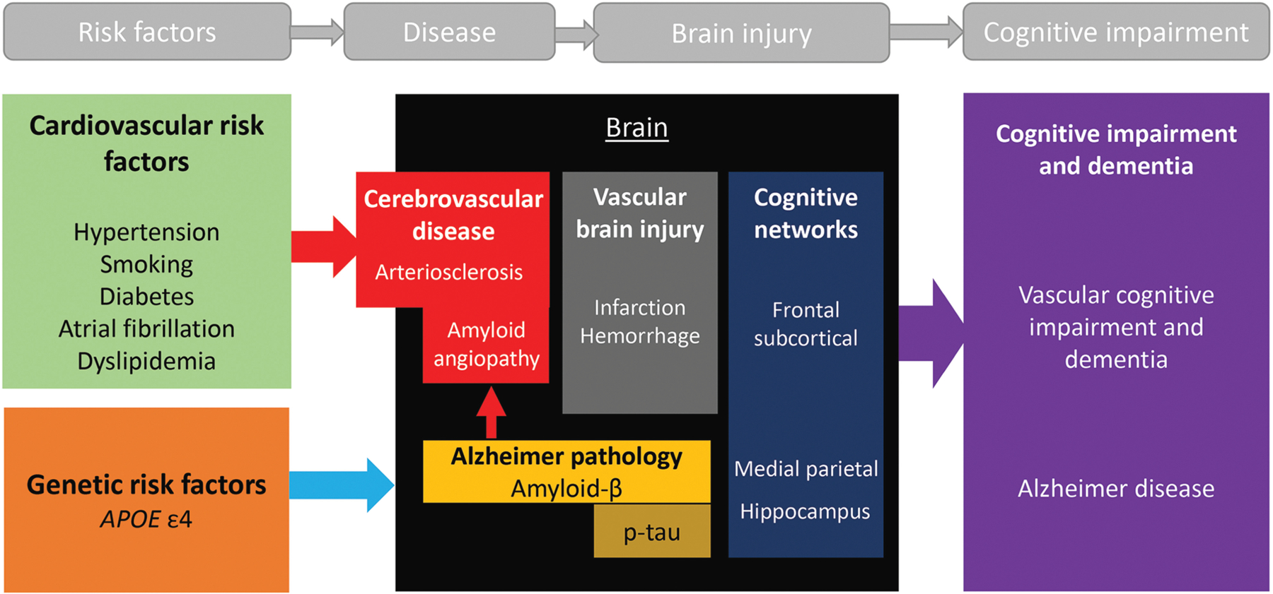
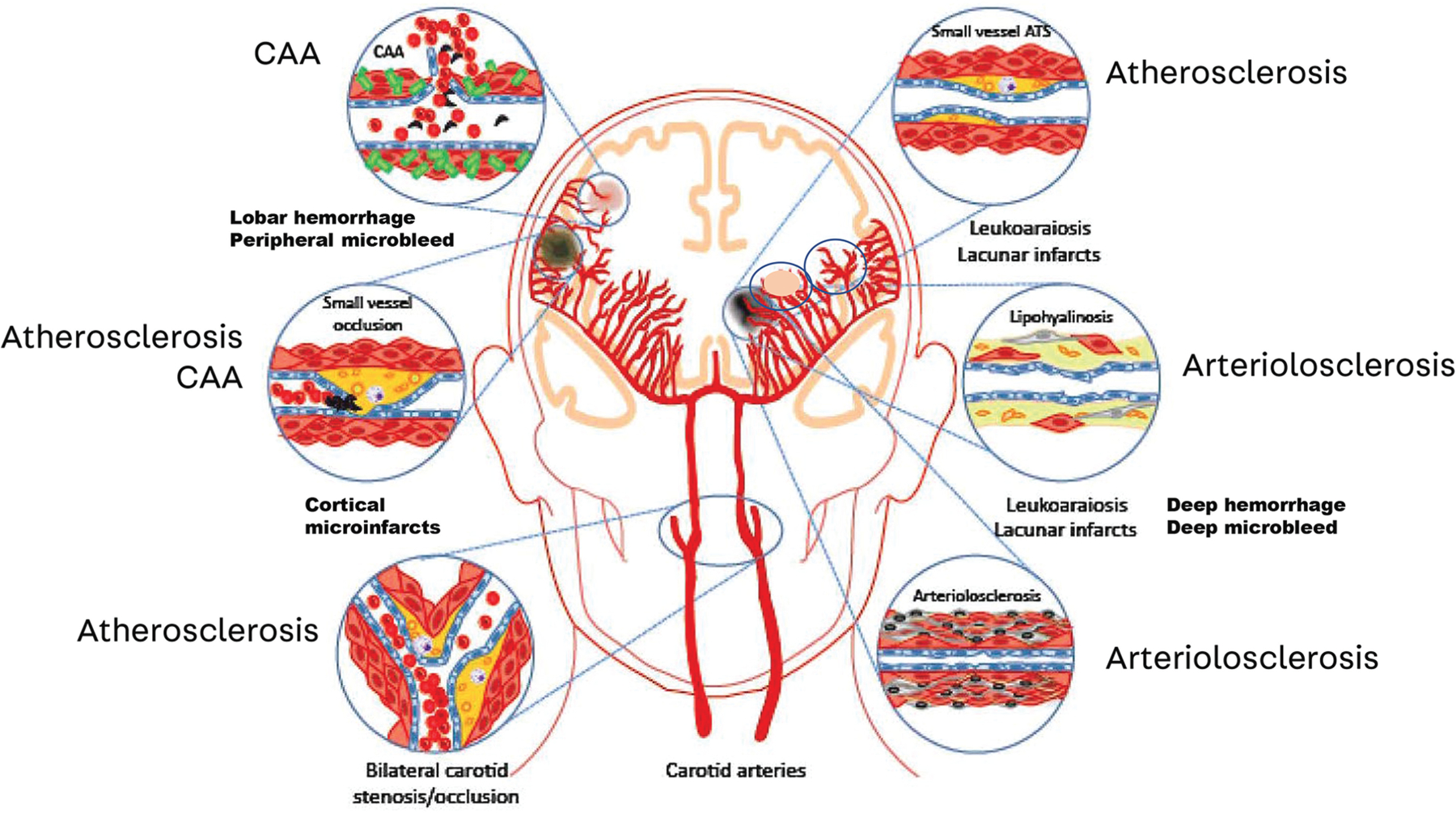
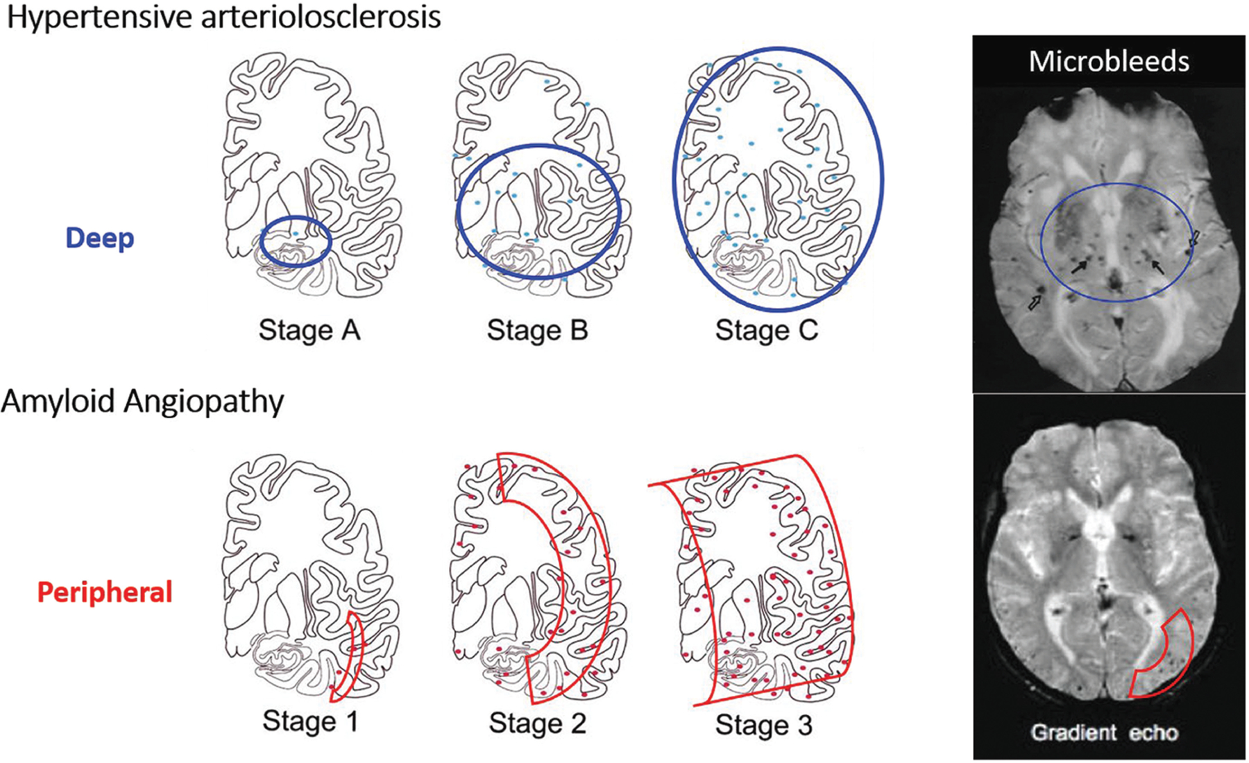
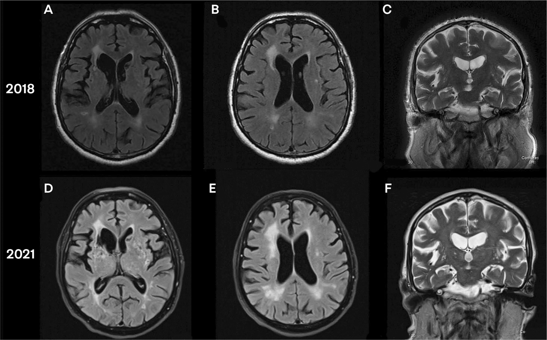
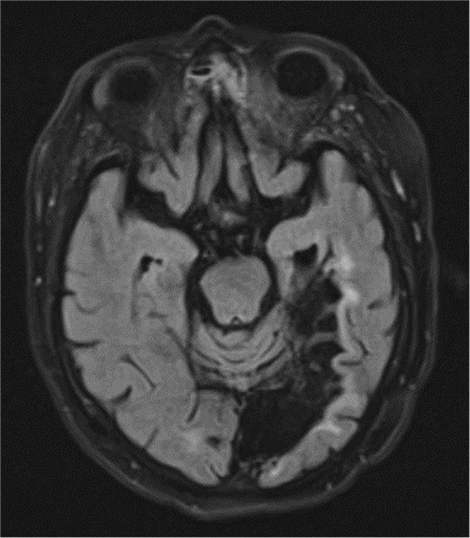
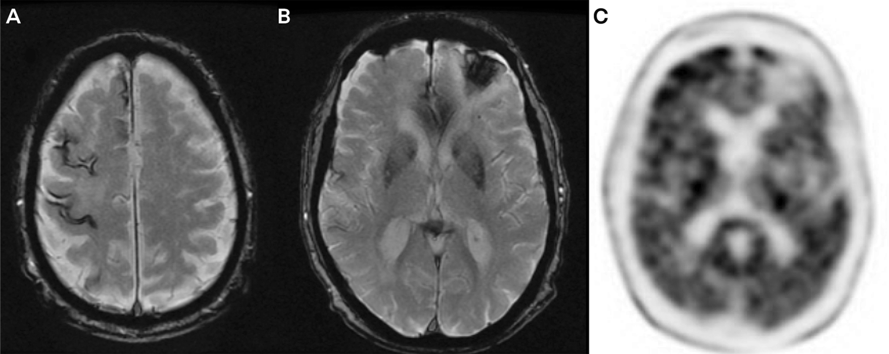
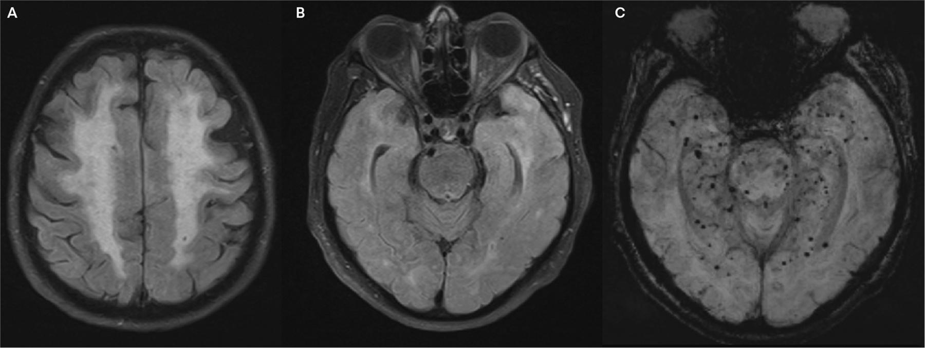
Purpose of review: This article gives a broad overview of vascular cognitive impairment and dementia, including epidemiology, pathophysiology, clinical approach, and management. Emphasis is placed on understanding the common underlying types of cerebrovascular disease (including atherosclerosis, arteriolosclerosis, and cerebral amyloid angiopathy) and awareness of rare inherited cerebrovascular disorders.

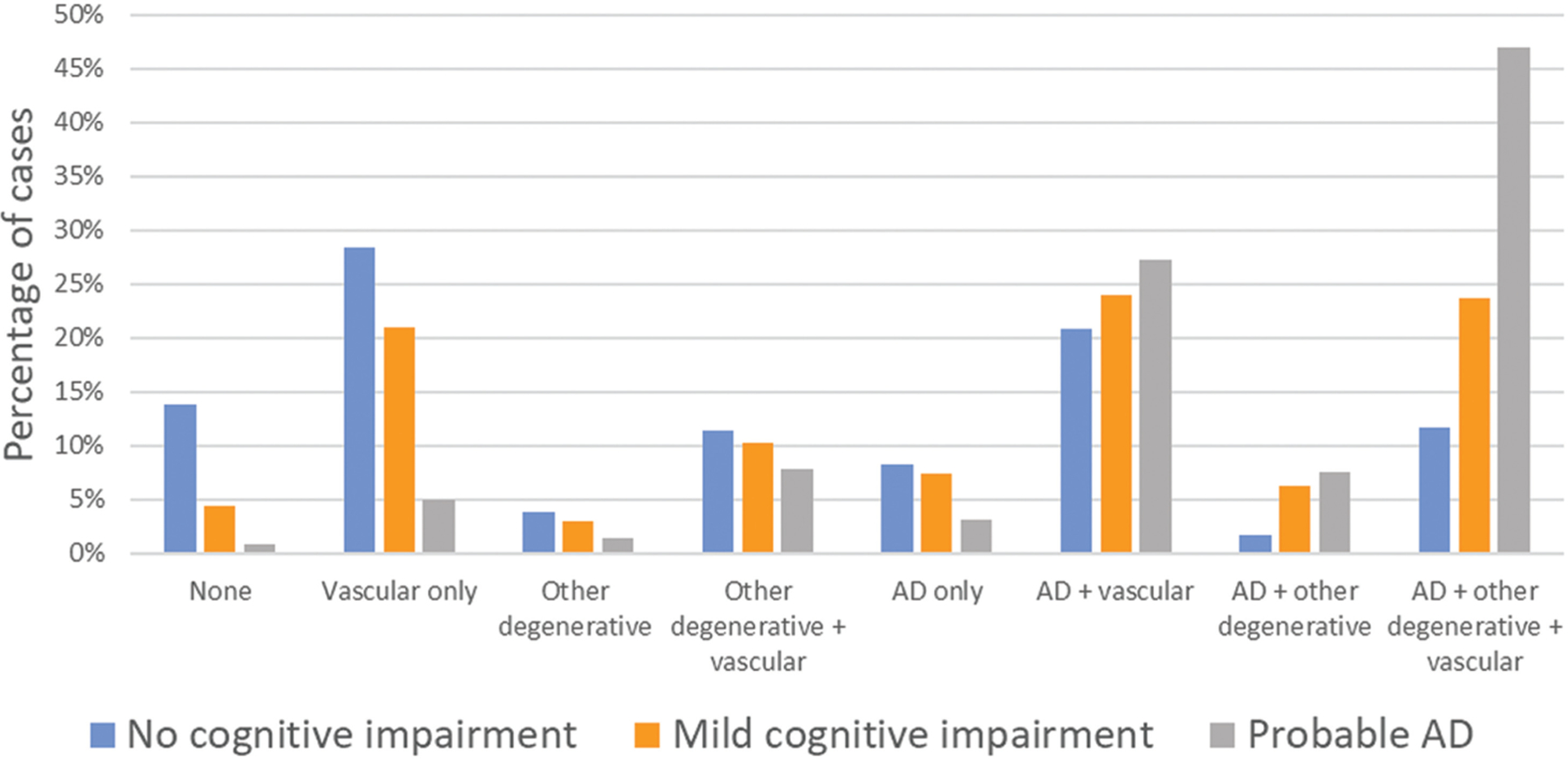
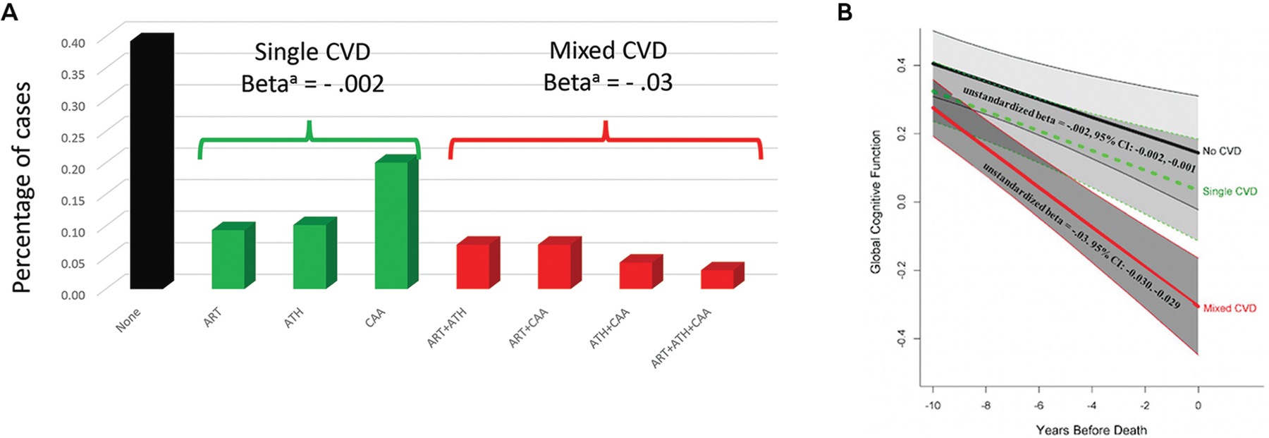
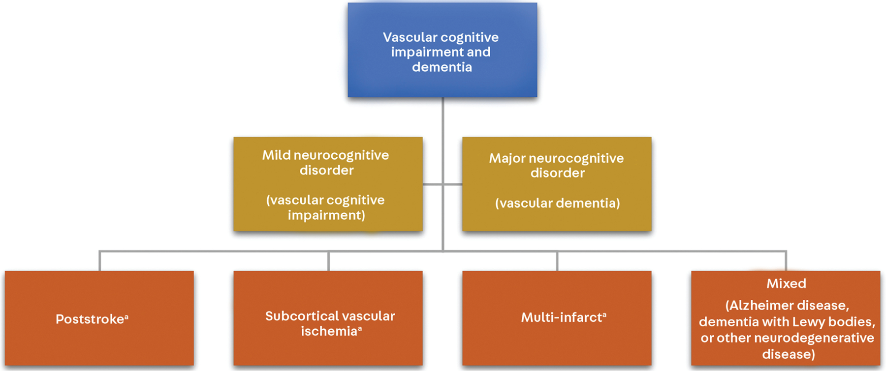
Recent findings: The pathophysiology of vascular cognitive impairment and dementia is heterogeneous, and the most recent diagnostic criteria for vascular cognitive impairment and dementia break down the diagnosis of major vascular dementia into four phenotypic categories, including subcortical ischemic vascular dementia, poststroke dementia, multi-infarct dementia, and mixed dementia. Control of cardiovascular risk factors, including management of midlife blood pressure, cholesterol, and blood sugars, remains the mainstay of prevention for vascular cognitive impairment and dementia. Cerebral amyloid angiopathy requires special consideration when it comes to risk factor management given the increased risk of spontaneous intracerebral hemorrhage. Recent trials suggest some improvement in global cognitive function in patients with vascular cognitive impairment and dementia with targeted cognitive rehabilitation.

Summary: Thorough clinical evaluation and neuroimaging form the basis for diagnosis. As vascular cognitive impairment and dementia is the leading nondegenerative cause of dementia, identifying risk factors and optimizing their management is paramount. Once vascular brain injury has occurred, symptomatic management should be offered and secondary prevention pursued.


Figures

FIGURE 5–1
FIGURE 5–1
Prevalence of different neuropathologies among the participants in the Rush Religious Orders Study/Memory and Aging Project (ROS/MAP) who clinically had no cognitive impairment, mild cognitive impairment, or probable Alzheimer disease (AD) (n = 1078). “Other degenerative” refers to other neurodegenerative pathology, including Lewy body, transactive response DNA-binding protein 43 (TDP-43) and hippocampal sclerosis. Notably, when compiled, mixed pathology increases from about 46% of participants with no cognitive impairment to 89.7% of participants with probable AD. a Data from Kapasi A, et al, Acta Neuropathologica.
FIGURE 5–2
FIGURE 5–2
Pathogenesis of vascular cognitive impairment and dementia and mixed Alzheimer disease/vascular cognitive impairment and dementia. Cardiovascular risk factors are the leading cause of atherosclerotic cardiovascular disease (ASCVD), which results in cerebral vascular disease and, uncontrolled, may result in vascular brain injury and ultimately vascular cognitive impairment and dementia. Apolipoprotein E4 (APOE ε4) is associated with Alzheimer disease (AD) and cerebral amyloid angiopathy (CAA) and is a frequent cause of mixed AD/vascular cognitive impairment and dementia. Vascular brain injury can result in damage to the frontal subcortical circuits, whereas AD tends to affect the medial parietal and hippocampal regions.
FIGURE 5–3
FIGURE 5–3
The different forms of cerebral vascular disease that may contribute to vascular cognitive impairment and dementia. ATS = atherosclerosis; CAA = cerebral amyloid angiopathy. Modified with permission from Iadecola C, Neuron. © 2013 Elsevier Inc.
FIGURE 5–4
FIGURE 5–4
The parts of the neurovascular unit. Reprinted with permission from Sweeney MD, et al, J Cereb Blood Flow Metab. © 2015 SAGE Publications.
FIGURE 5–5
FIGURE 5–5
The association of single (green) and mixed (red) cerebrovascular disease (CVD) profile groups with global cognitive decline compared with participants with no cerebrovascular disease (black). In an autopsy series on participants with Alzheimer disease from the Rush Religious Orders Study, Rush Memory and Aging Project, and the Minority Aging Research Study, the major types of cerebrovascular disease (CVD) and their various combinations were examined. Notably, mixed CVD produced greater cognitive decline over time when compared to single or no CVD groups. a Unstandardized beta for rate of cognitive decline for single CVD and mixed CVD compared to reference group with no CVD. ART = arteriolosclerosis; ATH = atherosclerosis; CAA = cerebral amyloid angiopathy. Panel B reprinted with permission from Lamar M, et al. © 2021 American Heart Association, Inc.
FIGURE 5–7
FIGURE 5–7
The VICCCS (Vascular Impairment of Cognition Classification Consensus Study) criteria for vascular cognitive impairment and dementia. a Although these subtypes are listed separately from “mixed,” any subtype of vascular cognitive impairment and dementia has the potential to have mixed pathology. Modified with permission from Skrobot O, et al, Alzheimers Dement. © 2016 The Alzheimer’s Association.
FIGURE 5–8
FIGURE 5–8
How infarct location is linked to cognitive function. The yellow to red coloration shows the level of statistical association between infarcts in those locations and cognitive function in multivariate region of interest-based analyses. Light blue corresponds to areas associated with cognition in univariate analyses. ACR = anterior corona radiata; AIC = anterior limb of internal capsule; BCC = body of corpus callosum; Cing = cingulum (white matter); EC = external capsule; GCC = genu of corpus callosum; IFGop = inferior frontal gyrus (opercular); IFGorb = inferior frontal gyrus (orbital); IFGtri = inferior frontal gyrus (triangular); IFOF = inferior fronto-occipital fasciculus; MOL = middle occipital lobe; MTG = middle temporal gyrus; MTP = middle temporal pole; PCR = posterior corona radiata; PIC = posterior limb of internal capsule; PTR = posterior thalamic radiation; RIC = retrolenticular part of internal capsule; SCR = superior corona radiata; SFOF = superior fronto-occipital fasciculus; SLF = superior longitudinal fasciculus; SPG = superior parietal gyrus; STG = superior temporal gyrus. Reprinted with permission from Zhao L, et al, J Cereb Blood Flow Metab. © 2018 SAGE Publications.
FIGURE 5–10
FIGURE 5–10
Visual reference for the Fazekas scale. Reprinted with permission from Inzitari D, et al, BMJ. © 2009 BMJ Publishing Group Ltd.
FIGURE 5–11
FIGURE 5–11
Distinguishing the location and staging the severity of microbleeds. Hypertensive arteriosclerosis generally results in deep microbleeds, indicated by blue ovals, whereas amyloid angiopathy is characterized by peripheral microbleeds, indicated by red shapes. Modified with permission from Thal DR, et al, J Neuropathol Exp Neurol. © 2003 Oxford University Press.
FIGURE 5–6
FIGURE 5–6
Imaging of the patient in CASE 5-1. A, Axial fluid-attenuated inversion recovery (FLAIR) MRI shows lacunar infarct of bilateral caudate and right external capsule from initial presentation. B, Axial FLAIR MRI shows white matter disease (Fazekas scale grade 2) from initial presentation. C, Coronal T2-weighted MRI shows hippocampal sparing from initial presentation. D, Axial FLAIR MRI shows progression of white matter disease and lacunar infarcts on left basal ganglia as well as adjacent to right anterior horn, 3 years later. E, Axial FLAIR MRI shows white matter disease (Fazekas scale grade 3), 3 years later. F, Coronal T2-weighted MRI shows continued hippocampal sparing, despite progression in generalized atrophy (slight enlargement of ventricles can be seen), 3 years later.
FIGURE 5–9
FIGURE 5–9
Imaging of the patient in CASE 5-2. Axial fluid-attenuated inversion recovery (FLAIR) MRI shows encephalomalacia in the left posterior cerebral artery territory as well as hippocampal atrophy, left greater than right.
FIGURE 5–12
FIGURE 5–12
Imaging of the patient in CASE 5-3. A, Axial gradient recalled echo (GRE) MRI shows superficial siderosis along the right parietal and right frontal sulci, consistent with cerebral amyloid angiopathy. B, Axial GRE MRI shows hemosiderin deposition in the left frontal lobe. C, Axial amyloid positron emission tomography (PET) shows amyloid positivity consistent with cerebral amyloid angiopathy and Alzheimer pathology.
FIGURE 5–13
FIGURE 5–13
Imaging of the patient in CASE 5-4. A, Axial fluid-attenuated inversion recovery (FLAIR) MRI shows severe confluent white matter disease consistent with Fazekas scale grade 3. B, Axial FLAIR MRI slice through the temporal lobes shows confluent white matter disease involving the temporal poles, left more than right. C, Axial gradient recalled echo (GRE) MRI shows innumerable microbleeds throughout the temporal lobes bilaterally.

Similar articles

Cited by

References

    1. American Psychiatric Association. Diagnostic and Statistical Manual of Mental Disorders 5th ed. American Psychiatric Association; 2013.
    1. Rizzi L, Rosset I, Roriz-Cruz M. Global epidemiology of dementia: Alzheimer’s and vascular types. Biomed Res Int 2014;2014(908915). doi:10.1155/2014/908915 - DOI - PMC - PubMed
    1. Willis T De anima brutorum quae hominis vitalis ac sentitiva est: exercitationes duae. Typis E.F. impensis Ric Davis, Oxon; 1672.
    1. Gorelick PB, Scuteri A, Black SE, et al. Vascular contributions to cognitive impairment and dementia: a statement for healthcare professionals from the American Heart Association/American Stroke Association. Stroke 2011;42(9):2672–2713. doi:10.1161/STR.0b013e3182299496 - DOI - PMC - PubMed
    1. Hachinski VC, Iliff LD, Zilhka E, et al. Cerebral blood flow in dementia. Arch Neurol 1975;32(9): 632–637. doi:10.1001/archneur.1975. 00490510088009 - DOI - PubMed

MeSH terms