

Mitochondria ROS and mitophagy in acute kidney injury
- PMID: 35678504
- PMCID: PMC9851232
- DOI: 10.1080/15548627.2022.2084862
Mitochondria ROS and mitophagy in acute kidney injury
Abstract
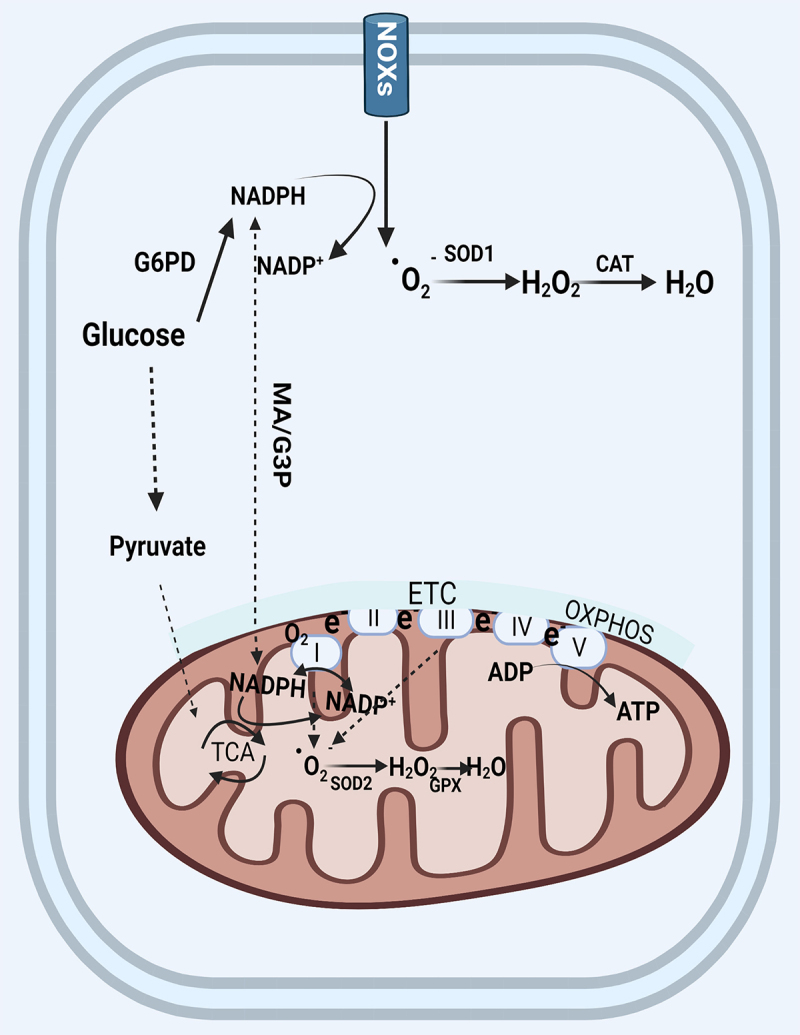
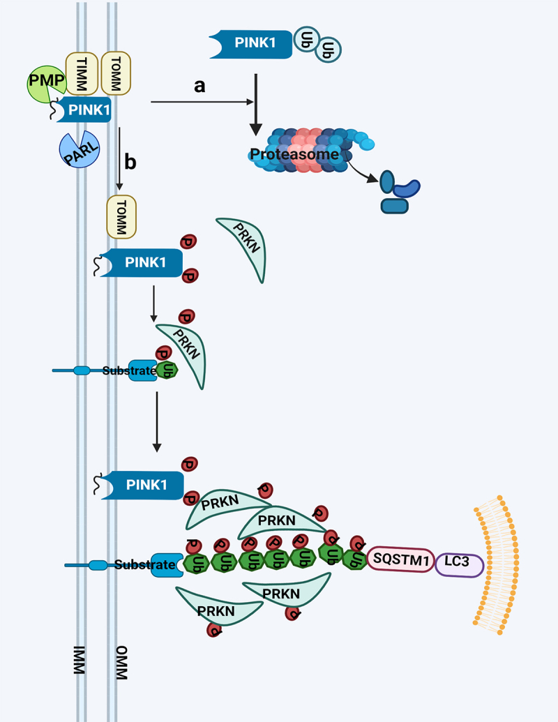
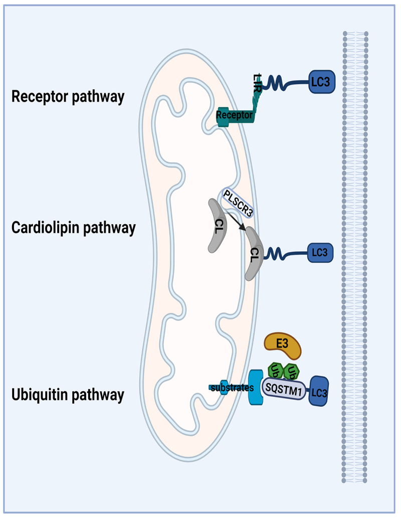
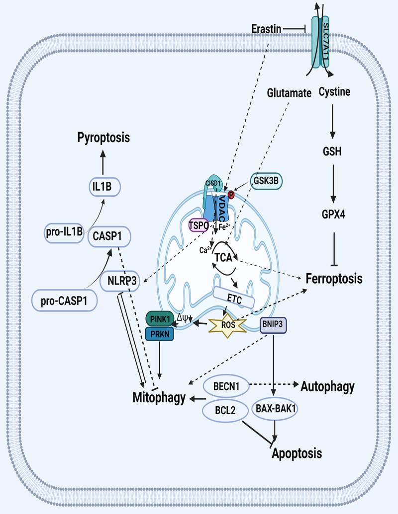
Mitophagy is an essential mitochondrial quality control mechanism that eliminates damaged mitochondria and the production of reactive oxygen species (ROS). The relationship between mitochondria oxidative stress, ROS production and mitophagy are intimately interwoven, and these processes are all involved in various pathological conditions of acute kidney injury (AKI). The elimination of damaged mitochondria through mitophagy in mammals is a complicated process which involves several pathways. Furthermore, the interplay between mitophagy and different types of cell death, such as apoptosis, pyroptosis and ferroptosis in kidney injury is unclear. Here we will review recent advances in our understanding of the relationship between ROS and mitophagy, the different mitophagy pathways, the relationship between mitophagy and cell death, and the relevance of these processes in the pathogenesis of AKI.Abbreviations: AKI: acute kidney injury; AMBRA1: autophagy and beclin 1 regulator 1; ATP: adenosine triphosphate; BAK1: BCL2 antagonist/killer 1; BAX: BCL2 associated X, apoptosis regulator; BCL2: BCL2 apoptosis regulator; BECN1: beclin 1; BH3: BCL2 homology domain 3; BNIP3: BCL2 interacting protein 3; BNIP3L/NIX: BCL2 interacting protein 3 like; CASP1: caspase 1; CAT: catalase; CCCP: carbonyl cyanide m-chlorophenylhydrazone; CI-AKI: contrast-induced acute kidney injury; CISD1: CDGSH iron sulfur domain 1; CL: cardiolipin; CNP: 2',3'-cyclic nucleotide 3'-phosphodiesterase; DNM1L/DRP1: dynamin 1 like; E3: enzyme 3; ETC: electron transport chain; FA: folic acid; FUNDC1: FUN14 domain containing 1; G3P: glycerol-3-phosphate; G6PD: glucose-6-phosphate dehydrogenase; GPX: glutathione peroxidase; GSH: glutathione; GSK3B: glycogen synthase kinase 3 beta; GSR: glutathione-disulfide reductase; HIF1A: hypoxia inducible factor 1 subunit alpha; HUWE1: HECT, UBA and WWE domain containing 1; IL1B: interleukin 1 beta; IMM: inner mitochondrial membrane; IPC: ischemic preconditioning; IRI: ischemia-reperfusion injury; LIR: LC3-interacting region; LPS: lipopolysaccharide; MA: malate-aspartate; MPT: mitochondrial permeability transition; MUL1: mitochondrial E3 ubiquitin protein ligase 1; mtROS: mitochondrial ROS; NLR: NOD-like receptor; NLRP3: NLR family pyrin domain containing 3; NOX: NADPH oxidase; OGD-R: oxygen-glucose deprivation-reperfusion; OMM: outer mitochondrial membrane; OPA1: OPA1 mitochondrial dynamin like GTPase; OXPHOS: oxidative phosphorylation; PARL: presenilin associated rhomboid like; PINK1: PTEN induced kinase 1; PLSCR3: phospholipid scramblase 3; PMP: peptidase, mitochondrial processing; PRDX: peroxiredoxin; PRKN: parkin RBR E3 ubiquitin protein ligase; RPTC: rat proximal tubular cells; ROS: reactive oxygen species; SLC7A11/xCT: solute carrier family 7 member 11; SOD: superoxide dismutase; SOR: superoxide reductase; SQSTM1/p62: sequestosome 1; TCA: tricarboxylic acid; TIMM: translocase of inner mitochondrial membrane; TOMM: translocase of outer mitochondrial membrane; TXN: thioredoxin; VDAC: voltage dependent anion channel; VCP: valosin containing protein.
Keywords: Acute kidney injury; cell death; mitochondria; mitophagy; reactive oxygen species.
Conflict of interest statement
None of the authors declared any conflict of interest in this work.
Figures
Similar articles
-
Clearance of damaged mitochondria via mitophagy is important to the protective effect of ischemic preconditioning in kidneys.Autophagy. 2019 Dec;15(12):2142-2162. doi: 10.1080/15548627.2019.1615822. Epub 2019 May 22. Autophagy. 2019. PMID: 31066324 Free PMC article.
-
Organelle-specific autophagy in inflammatory diseases: a potential therapeutic target underlying the quality control of multiple organelles.Autophagy. 2021 Feb;17(2):385-401. doi: 10.1080/15548627.2020.1725377. Epub 2020 Feb 12. Autophagy. 2021. PMID: 32048886 Free PMC article. Review.
-
Emerging views of mitophagy in immunity and autoimmune diseases.Autophagy. 2020 Jan;16(1):3-17. doi: 10.1080/15548627.2019.1603547. Epub 2019 Apr 21. Autophagy. 2020. PMID: 30951392 Free PMC article.
-
The multifaceted regulation of mitophagy by endogenous metabolites.Autophagy. 2022 Jun;18(6):1216-1239. doi: 10.1080/15548627.2021.1975914. Epub 2021 Sep 29. Autophagy. 2022. PMID: 34583624 Free PMC article.
-
Role of AMBRA1 in mitophagy regulation: emerging evidence in aging-related diseases.Autophagy. 2024 Dec;20(12):2602-2615. doi: 10.1080/15548627.2024.2389474. Epub 2024 Sep 2. Autophagy. 2024. PMID: 39113560 Free PMC article. Review.
Cited by
-
Apoptosis, Mitochondrial Autophagy, Fission, and Fusion Maintain Mitochondrial Homeostasis in Mouse Liver Under Tail Suspension Conditions.Int J Mol Sci. 2024 Oct 18;25(20):11196. doi: 10.3390/ijms252011196. Int J Mol Sci. 2024. PMID: 39456978 Free PMC article.
-
LONP1 alleviates ageing-related renal fibrosis by maintaining mitochondrial homeostasis.J Cell Mol Med. 2024 Sep;28(17):e70090. doi: 10.1111/jcmm.70090. J Cell Mol Med. 2024. PMID: 39261902 Free PMC article.
-
Dendrobium officinale Polysaccharides as a Natural Functional Component for Acetic-Acid-Induced Gastric Ulcers in Rats.Molecules. 2024 Feb 16;29(4):880. doi: 10.3390/molecules29040880. Molecules. 2024. PMID: 38398633 Free PMC article.
-
Regulation of ULK1 by WTAP/IGF2BP3 axis enhances mitophagy and progression in epithelial ovarian cancer.Cell Death Dis. 2024 Jan 29;15(1):97. doi: 10.1038/s41419-024-06477-0. Cell Death Dis. 2024. PMID: 38286802 Free PMC article.
-
Transcriptomics analysis revealed that TAZ regulates the proliferation of KIRC cells through mitophagy.BMC Cancer. 2024 Feb 19;24(1):229. doi: 10.1186/s12885-024-11903-9. BMC Cancer. 2024. PMID: 38373978 Free PMC article.
References
-
- Xiong H, Chen S, Lai L, et al. Modulation of miR-34a/SIRT1 signaling protects cochlear hair cells against oxidative stress and delays age-related hearing loss through coordinated regulation of mitophagy and mitochondrial biogenesis. Neurobiol Aging. 2019;79:30–42. - PubMed
Publication types
MeSH terms
Substances
Grants and funding
LinkOut - more resources
Full Text Sources
Research Materials
Miscellaneous