. 2022 Jun 29;10(3):e0020222.
doi: 10.1128/spectrum.00202-22. Epub 2022 Jun 9.

The CRISPR-Cas System Differentially Regulates Surface-Attached and Pellicle Biofilm in Salmonella enterica Serovar Typhimurium

Affiliations

The CRISPR-Cas System Differentially Regulates Surface-Attached and Pellicle Biofilm in Salmonella enterica Serovar Typhimurium

Nandita Sharma et al. Microbiol Spectr. .

Abstract

The CRISPR-Cas mediated regulation of biofilm by Salmonella enterica serovar Typhimurium was investigated by deleting CRISPR-Cas components ΔcrisprI, ΔcrisprII, ΔΔcrisprI crisprII, and Δcas op. We determined that the system positively regulates surface biofilm while inhibiting pellicle biofilm formation. Results of real-time PCR suggest that the flagellar (fliC, flgK) and curli (csgA) genes were repressed in knockout strains, causing reduced surface biofilm. The mutants displayed altered pellicle biofilm architecture. They exhibited bacterial multilayers and a denser extracellular matrix with enhanced cellulose and less curli, ergo weaker pellicles than those of the wild type. The cellulose secretion was more in the knockout strains due to the upregulation of bcsC, which is necessary for cellulose export. We hypothesized that the secreted cellulose quickly integrates into the pellicle, leading to enhanced pellicular cellulose in the knockout strains. We determined that crp is upregulated in the knockout strains, thereby inhibiting the expression of csgD and, hence, also of csgA and bcsA. The conflicting upregulation of bcsC, the last gene of the bcsABZC operon, could be caused by independent regulation by the CRISPR-Cas system owing to a partial match between the CRISPR spacers and bcsC gene. The cAMP-regulated protein (CRP)-mediated regulation of the flagellar genes in the knockout strains was probably circumvented through the regulation of yddx governing the availability of the sigma factor σ28 that further regulates class 3 flagellar genes (fliC, fljB, and flgK). Additionally, the variations in the lipopolysaccharide (LPS) profile and expression of LPS-related genes (rfaC, rfbG, and rfbI) in knockout strains could also contribute to the altered pellicle architecture. Collectively, we establish that the CRISPR-Cas system differentially regulates the formation of surface-attached and pellicle biofilm. IMPORTANCE In addition to being implicated in bacterial immunity and genome editing, the CRISPR-Cas system has recently been demonstrated to regulate endogenous gene expression and biofilm formation. While the function of individual cas genes in controlling Salmonella biofilm has been explored, the regulatory role of CRISPR arrays in biofilm is less studied. Moreover, studies have focused on the effects of the CRISPR-Cas system on surface-associated biofilms, and comprehensive studies on the impact of the system on pellicle biofilm remain an unexplored niche. We demonstrate that the CRISPR array and cas genes modulate the expression of various biofilm genes in Salmonella, whereby surface and pellicle biofilm formation is distinctively regulated.

Keywords: Salmonella; pellicle biofilm; surface-attached biofilm; type I-E CRISPR-Cas system.


Conflict of interest statement

The authors declare no conflict of interest.

Figures

FIG 1
FIG 1
The CRISPR-Cas system knockout strains of S. enterica subsp. enterica serovar Typhimurium 14028s showed reduced biofilm formation under gallstone-mimicking conditions (A), while these strains showed temporal variations in biofilm at the solid-liquid-air interface (B). (A) Wild-type, CRISPR, and cas operon knockout strains transformed with empty vector pQE60 (WT60, ΔcrisprI 60, ΔcrisprII 60, Δcas op. 60, and ΔΔcrisprI crisprII 60), and the complement strains (ΔcrisprI+pcrisprI and ΔcrisprII+pcrisprII) were cultured in cholesterol-coated microcentrifuge tubes in LB media for 96 h at 37°C under static conditions. (B) S. Typhimurium strain 14028s wild-type (WT), CRISPR (ΔcrisprI, ΔcrisprII, and ΔΔcrisprI crisprII) and cas operon (Δcas op.) knockout strains were cultured in LB without NaCl media for different time periods (24 h, 48 h, and 96 h) at 25°C under static conditions. Biofilm formation was estimated using the crystal violet staining method. Graph represents optical density at 570 nm (OD570) for each strain, normalized by OD570 of WT. An unpaired t test was used to determine significant differences between the WT and knockout strains. Error bars indicate SD. Statistical significance is shown as follows: *, P ≤ 0.05; **, P ≤ 0.01; ***, P ≤ 0.001; ****, P < 0.0001; and ns, not significant. A.U., arbitrary units.
FIG 2
FIG 2
Morphology of the air-exposed side (A) and liquid-submerged side (B) of pellicle biofilm at 96 h. The strains were grown in LB without NaCl for 96 h at 25°C under static conditions. The pellicle biofilms were fixed using 2.5% glutaraldehyde and dehydrated with increasing ethanol concentrations. SEM image analysis depicts the difference in the pellicle biofilm architecture of CRISPR-Cas knockout (ΔcrisprI, ΔcrisprII, Δcas op., and ΔΔcrisprI crisprII) strains and that of the wild type (WT) for both the air-exposed side (captured at ×10,000 magnification) and liquid-submerged side (captured at ×2,500 magnification) of pellicle biofilm. The air-exposed surface of the pellicle biofilm of CRISPR-Cas knockout strains had a denser, mat-like ECM. It consisted of “hilly” structures (marked with arrowheads), indicating more layering of the biofilm. The liquid-submerged surface of the pellicle biofilm of CRISPR-Cas knockout strains had more EPS lumps (marked with arrowheads) than the wild type. Images were scaled to bar.
FIG 3
FIG 3
Reduced swarming motility (A) and expression of the flagellar protein, FliC (B), was observed in the CRISPR-Cas system knockout strains. (A) Overnight cultures were point inoculated on swarm agar plates and incubated at 37°C for 9 h. Swarming rates (cm/h) of the wild-type (WT) strain, the knockout strains (ΔcrisprI, ΔcrisprII, Δcas op., and ΔΔcrisprI crisprII), and the complement strains (ΔcrisprI + pcrisprI, ΔcrisprII+ pcrisprII) were calculated. Graph represents the swarming rate relative to that of WT. (B) Strains were grown in LB without NaCl for different time periods (12 h, 24 h, and 96 h) at 25°C under static conditions. The expression of the flagellar protein in planktonic bacteria (B) at early time points (12 h and 24 h) and in pellicle biofilm (B) at a late time point (96 h) was assessed using Western blot analysis with antibodies against FliC. Even at higher protein concentration, FliC was not detected in the blot for pellicle sample of any strain, indicating repression of FliC expression in pellicle biofilm. ΔfliC was used as a negative control. An unpaired t test was used to determine significant differences between the WT and knockout strains. Error bar indicates SD. Statistical significance is represented as follows: *, P ≤ 0.05; **, P ≤ 0.01; ***, P ≤ 0.001; ****, P < 0.0001; ns, not significant. A.U., arbitrary units; #, ratio above the immunoblots (B), indicates the relative intensity of the bands with respect to wild type, observed in the blots, normalized by the relative intensity of the bands with respect to wild type, observed on the gel. The ratio is as follows: (FliC intensity/Coomassie band intensity)strain(FliC intensity/Coomassie band intensity)WT.
FIG 4
FIG 4
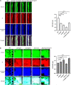
The CRISPR-Cas knockout strains showed temporal variations in their bacterial cell concentration, cellulose content, and SYTO9/PI ratio compared to WT at early (24 h) (A) and late (96 h) time points (B). (A and B) The S. Typhimurium strain 14028s wild-type (WT), CRISPR (ΔcrisprI, ΔcrisprII, and ΔΔcrisprI crisprII), and cas operon (Δcas op.) knockout strains were cultured in LB without NaCl for 24 h (A) and 96 h (B) at 25°C under static conditions. The biofilm formed was stained with SYTO9, propidium iodide (PI), and calcofluor white for 30 min in the dark at RT. The CLSM images were captured, and orthogonal projections of wild-type and CRISPR-Cas knockout strains were obtained. Graphs on the right of the CLSM images represent the ratio of mean intensity of SYTO9 to mean intensity of PI for respective strains at 24 h (A) and 96 h (B). An unpaired t test was used to determine significant differences between the WT and knockout strains. Error bars indicate SD. SD. Statistical significance is shown as follows: *, P ≤ 0.05; **, P ≤ 0.01; ***, P ≤ 0.001; ****, P < 0.0001; and ns, not significant. A.U., arbitrary units.
FIG 5
FIG 5
CRISPR-Cas knockout strains showed variations in the production of key components like curli (A) and cellulose (B and C). Curli production in the planktonic culture and pellicle biofilms of wild-type, CRISPR, and cas operon knockout strains was assessed with the help of Congo red depletion. The S. Typhimurium strain 14028s wild-type (WT), CRISPR (ΔcrisprI, ΔcrisprII, and ΔΔcrisprI crisprII) and cas operon (Δcas op.) knockout strains were cultured in LB without NaCl for different time periods (24 h and 96 h) at 25°C under static conditions. (A) Congo red depletion was determined by measuring absorbance at 500 nm. Graph represents absorbance at 500 nm of each strain, normalized by absorbance at 500 nm of WT. (B) Cellulose production in the pellicle biofilms of wild-type, CRISPR, and cas operon knockout strains was quantitatively assessed by determining the cellulose dry weight in the pellicle biofilm. The S. Typhimurium strain 14028s WT, CRISPR (ΔcrisprI, ΔcrisprII, and ΔΔcrisprI crisprII) and cas operon (Δcas op.) knockout strains were cultured in LB without NaCl for different time periods (48 h and 96 h) at 25°C under static conditions. (C) Qualitative analysis of the amount of cellulose present in the biofilm was done by measuring the calcofluor intensity of the CLSM images (represented in Fig. 4A and B). The S. Typhimurium strain 14028s WT, CRISPR (ΔcrisprI, ΔcrisprII, and ΔΔcrisprI crisprII), and cas operon (Δcas op.) knockout strains were cultured in LB without NaCl 96 h at 25°C under static conditions. An unpaired t test was used to determine significant differences between the WT and knockout strains. Error bar indicates SD. Statistical significance is shown as follows: *, P ≤ 0.05; **, P ≤ 0.01; ***, P ≤ 0.001; ****, P < 0.0001; and ns, not significant. A.U., arbitrary units.
FIG 6
FIG 6
CRISPR-Cas system knockout strains showed differences in the expressions of flagellar genes (A), the production of curli-csgA (B) curli-csgD (C), LPS-rfaC (D), cellulose-bcsC (E), and cAMP-regulated protein (crp) (F) compared to WT. S. Typhimurium strain 14028s wild-type (WT), CRISPR (ΔcrisprI, ΔcrisprII and ΔcrisprI ΔcrisprII), and cas operon (Δcas op.) knockout strains were cultured in LB without NaCl for different time periods (24 h and 96 h) at 25°C under static conditions. Total RNA was isolated from bacteria (24 h) and pellicle biofilm (96 h). One microgram of RNA was used for cDNA synthesis, followed by qRT-PCR. Relative expression of the gene was calculated using the 2−ΔΔCT method and normalized to reference gene, rpoD.
FIG 7
FIG 7
Differential regulation of surface-attached and pellicle biofilm formation in S. Typhimurium by the CRISPR-Cas system.

Similar articles

Cited by

References

    1. Pinilla-Redondo R, Shehreen S, Marino ND, Fagerlund RD, Brown CM, Sørensen SJ, Fineran PC, Bondy-Denomy J. 2020. Discovery of multiple anti-CRISPRs highlights anti-defense gene clustering in mobile genetic elements. Nat Commun 11. doi:10.1038/s41467-020-19415-3. - DOI - PMC - PubMed
    1. Hille F, Charpentier E. 2016. CRISPR-Cas: biology, mechanisms and relevance. Philos Trans R Soc B 371:20150496. doi:10.1098/rstb.2015.0496. - DOI - PMC - PubMed
    1. Sampson TR, Weiss DS. 2014. CRISPR-Cas systems: new players in gene regulation and bacterial physiology. Front Cell Infect Microbiol 4:37. doi:10.3389/fcimb.2014.00037. - DOI - PMC - PubMed
    1. Bozic B, Repac J, Djordjevic M. 2019. Endogenous gene regulation as a predicted main function of type I-E CRISPR/Cas system in E. coli. Molecules 24:784. doi:10.3390/molecules24040784. - DOI - PMC - PubMed
    1. Shariat N, Timme RE, Pettengill JB, Barrangou R, Dudley EG. 2015. Characterization and evolution of Salmonella CRISPR-Cas systems. Microbiology (Reading) 161:374–386. doi:10.1099/mic.0.000005. - DOI - PubMed

Publication types