. 2022 Apr 30;44(5):1995-2014.
doi: 10.3390/cimb44050135.

Cold Atmospheric Plasma-Activated Media Improve Paclitaxel Efficacy on Breast Cancer Cells in a Combined Treatment Model

Affiliations

Cold Atmospheric Plasma-Activated Media Improve Paclitaxel Efficacy on Breast Cancer Cells in a Combined Treatment Model

Cosmin-Teodor Mihai et al. Curr Issues Mol Biol. .

Abstract

The use of plasma-activated media (PAM), an alternative to direct delivery of cold atmospheric plasma to cancer cells, has recently gained interest in the plasma medicine field. Paclitaxel (PTX) is used as a chemotherapy of choice for various types of breast cancers, which is the leading cause of mortality in females due to cancer. In this study, we evaluated an alternative way to improve anti-cancerous efficiency of PTX by association with PAM, the ultimate achievement being a better outcome in killing tumoral cells at smaller doses of PTX. MCF-7 and MDA-MB-231 cell lines were used, and the outcome was measured by cell viability (MTT assay), the survival rate (clonogenic assay), apoptosis occurrence, and genotoxicity (COMET assay). Treatment consisted of the use of PAM in combination with under IC50 doses of PTX in short- and long-term models. The experimental data showed that PAM had the capacity to improve PTX's cytotoxicity, as viability of the breast cancer cells dropped, an effect maintained in long-term experiments. A higher frequency of apoptotic, dead cells, and DNA fragmentation was registered in cells treated with the combined treatment as compared with those treated only with PT. Overall, PAM had the capacity to amplify the anti-cancerous effect of PTX.

Keywords: DNA damage; PTX; apoptosis; breast cancer cell; cell cytotoxicity; combined therapy; plasma-activated media.


Conflict of interest statement

The authors declare no conflict of interest. The funders had no role in the design of the study; in the collection, analyses, or interpretation of data; in the writing of the manuscript, or in the decision to publish the results.

Figures

Figure 1
Figure 1
The schema (A) and working setup (B) for air DBD plasma experimental exposure system for DMEM activation.
Figure 2
Figure 2
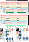
(A) Viability of the MCF-7 and MDA-MB-231 cell lines was determined by MTT method 24 and 48 h after the treatment with PTX in a dose ranging from 0.1 μM to 0.001 μM, PAM (30 s and 60 s), and combined treatment (PTX and PAM). PAM was obtained through media exposure to CAP for 30 s and 60 s. Treatment with PAM and incubation of the cells with PAM was 20 min. PTX was applied for 24 h and combined treatment consisted of PAM application followed by PTX incubation. Cell viability was determined by MTT assay. Control group received only normal media; PAM—treatment was applied only with plasma-activated media; PTX—treatment was applied only with PTX; PTX and PAM—combined treatment was used. Data are mean and SEM values (n = 8). (B) Statistical significance obtained by multiple comparison between different groups. Colored dots indicate the statistical significance computed after applying Tukey multiple comparison test and are in accordance with the obtained value: darker values correspond to p < 0.0001 and lighter ones to non-significant values.
Figure 3
Figure 3
Representative photographs of the MCF-7 and MDA-MB-231 colonies and mean values of the survival fractions as calculated in the case of the control group, PAM 15 s, and PAM 30 s, 0.01 µM PTX, combined treatment (0.01 µM PTX and PAM 15 s and 0.01 µM PTX and PAM 30 s). Data are mean and SEM values (n = 3). * p < 0.05; *** p < 0.001 (ANOVA).
Figure 4
Figure 4
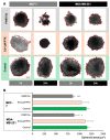
Spheroid area assessment of the MCF-7 and MDA-MB-231 cell culture experimental variants. (A) Representative images of the MCF-7 and MDA-MB-231 spheroids obtained by hanging drop method and treated with PTX and PAM 15 s. Column 0 h represents the spheroids before the treatment was added, while column 24 h represents the images of spheroids 24 h after the treatment. The evaluation of shrinkage/growth was assessed by taking photographs of the same spheroid as at the beginning of the treatment. Analysis consisted of the calculation of the area of total spheroid by Fiji software. (B) Mean area of spheroids (pixels2) following the treatment with PTX and PAM 15 s were acquired at 24 h after the treatment. Data are mean and SEM values (n = 8). * p < 0.05; ** p < 0.01; *** p < 0.001 (ANOVA).
Figure 5
Figure 5
Apoptosis assessment of MCF-7 and MDA-MB-231 cells treated with PAM and PTX. (A) Representative images of MCF-7 and MDA-MB-231 under the effect of PTX, combined treatment, and PAM. Untreated cells were cultivated in normal DMEM, without addition of any treatment. PTX-treated cells presented changes in morphology and an increased frequency of apoptotic (condensed green nucleus or orange chromatin) and dead cells. In 0.01 µM PTX and PAM 15 s and 0.01 µM PTX and PAM 30 s groups, the frequency of apoptotic and dead cells significantly increased. PAM 15 s and PAM 30 s showed less frequency of apoptotic and dead cells. (B) Distribution of the live, dead, and apoptotic (early and late apoptotic cells) populations (%) of MCF-7 and MDA-MB-231 cell cultures. Data are mean and SEM values (n = 3). * p < 0.05; ** p < 0.01; *** p < 0.001 (ANOVA).
Figure 6
Figure 6
(A) Effect of PAM, PTX, and combined treatment on cell migration of MCF-7 and MDA-MB-231 cells, representative images (all images have the same scale as shown in MCF-7 control image, 500 μm). Confluent layers of both cell lines were scratched, and the growth media were exchanged with corresponding media containing PTX and PTX and PAM, while for the control group only fresh complete media were added. Migration of MCF-7 and MDA-MB-231 cells was assessed by taking photos in the same spot at 24 and 48 h (n = 3) after the treatment was applied. The baseline was chosen as the initial area delimited by the margins of the scratch, before the treatment was added. The 0.01 µM PTX and PAM 15 s and 0.01 µM PTX and PAM 30 s significantly decreased migration to 48 h (p < 0.05, p < 0.01). (B) The computed results are presented as mean ± SEM values for 3 independent tests (n = 3). The statistical significance was established by comparative analysis of the treated groups against PTX using one-way analysis of variance followed by the Tukey post hoc test. * p < 0.05, ** p < 0.01, *** p < 0.001.
Figure 7
Figure 7
Evaluation of genotoxicity in MCF-7 and MDA-MB-231 cells. (A,D) Relative frequency of DNA content in experimental groups; (B,E) content of DNA (%) COMET head; (C,F) and characteristic microscopic photos of comets, indicating the genotoxic impact of the treatment applied with PTX, PAM, or combined treatment. Data are mean and SEM values (n = 3). One-way analysis of variance followed by the Tukey post hoc test was applied in order to evaluate the statistical significance. * p < 0.05, ** p < 0.01, *** p < 0.001.

References

    1. Bray F., Ferlay J., Soerjomataram I., Siegel R.L., Torre L.A., Jemal A. Global Cancer Statistics 2018: GLOBOCAN Estimates of Incidence and Mortality Worldwide for 36 Cancers in 185 Countries. CA. Cancer J. Clin. 2018;68:394–424. doi: 10.3322/caac.21492. - DOI - PubMed
    1. Gerber B., Müller H., Reimer T., Krause A., Friese K. Nutrition and Lifestyle Factors on the Risk of Developing Breast Cancer. Breast Cancer Res. Treat. 2003;79:265–276. doi: 10.1023/A:1023959818513. - DOI - PubMed
    1. Gillet J.-P., Gottesman M.M. Mechanisms of Multidrug Resistance in Cancer. In: Zhou J., editor. Multi-Drug Resistance in Cancer. Volume 596. Humana Press; Totowa, NJ, USA: 2010. pp. 47–76. Methods in Molecular Biology. - PubMed
    1. Liu Y., Tan S., Zhang H., Kong X., Ding L., Shen J., Lan Y., Cheng C., Zhu T., Xia W. Selective Effects of Non-Thermal Atmospheric Plasma on Triple-Negative Breast Normal and Carcinoma Cells through Different Cell Signaling Pathways. Sci. Rep. 2017;7 doi: 10.1038/s41598-017-08792-3. - DOI - PMC - PubMed
    1. Delude C.M. Expensive Cancer Therapies: Unintended Effects. J. Natl. Cancer Inst. 2015;107:497. doi: 10.1093/jnci/dju497. - DOI - PubMed

LinkOut - more resources