

Sostdc1 Secreted from Cutaneous Lymphatic Vessels Acts as a Paracrine Factor for Hair Follicle Growth
- PMID: 35678675
- PMCID: PMC9164032
- DOI: 10.3390/cimb44050146
Sostdc1 Secreted from Cutaneous Lymphatic Vessels Acts as a Paracrine Factor for Hair Follicle Growth
Abstract
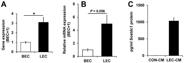
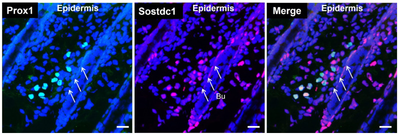
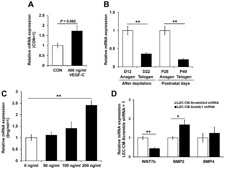
In our previous study, we found that lymphatic vessels stimulate hair follicle growth through paracrine effects on dermal papilla cells. However, the paracrine factors secreted from cutaneous lymphatic vessels that can activate dermal papilla cells are still unknown. In this study, we investigated whether lymphatic endothelial cells might secrete paracrine factors that activate dermal papilla cells in vitro. We found that Sostdc1 was more expressed in lymphatic endothelial cells compared with blood vascular endothelial cells. In addition, Sostdc1 expression levels were significantly increased during the anagen phase in the back skin of C57BL/6J mice, as compared to the telogen phase. We also observed that incubation of dermal papilla cells with 200 ng/mL Sostdc1 for 72 h induced the expression levels of Lef-1, a downstream target of Wnt signaling. Taken together, our results reveal that Sostdc1, a BMP antagonist, secreted from cutaneous lymphatic vessels, may act as a paracrine factor for hair follicle growth.
Keywords: Sostdc1; dermal papilla cells; hair follicle growth; lymphatic vessels.
Conflict of interest statement
The authors declare no conflict of interest.
Figures
Similar articles
-
Inhibitory autocrine factors produced by the mesenchyme-derived hair follicle dermal papilla may be a key to male pattern baldness.Br J Dermatol. 2006 Apr;154(4):609-18. doi: 10.1111/j.1365-2133.2006.07144.x. Br J Dermatol. 2006. PMID: 16536801
-
An important role of cutaneous lymphatic vessels in coordinating and promoting anagen hair follicle growth.PLoS One. 2019 Jul 25;14(7):e0220341. doi: 10.1371/journal.pone.0220341. eCollection 2019. PLoS One. 2019. PMID: 31344105 Free PMC article.
-
Immortalized rat whisker dermal papilla cells cooperate with mouse immature hair follicle buds to activate type IV procollagenases in collagen matrix coculture: correlation with ability to promote hair follicle development in nude mouse grafts.J Invest Dermatol. 1995 Aug;105(2):177-83. doi: 10.1111/1523-1747.ep12317089. J Invest Dermatol. 1995. PMID: 7636298
-
Do androgens influence hair growth by altering the paracrine factors secreted by dermal papilla cells?Eur J Dermatol. 2001 Jul-Aug;11(4):315-20. Eur J Dermatol. 2001. PMID: 11399537 Review.
-
Maintaining Hair Inductivity in Human Dermal Papilla Cells: A Review of Effective Methods.Skin Pharmacol Physiol. 2020;33(5):280-292. doi: 10.1159/000510152. Epub 2020 Oct 14. Skin Pharmacol Physiol. 2020. PMID: 33053562 Review.
Cited by
-
Genome-wide DNA methylation and transcriptome analyses reveal the key gene for wool type variation in sheep.J Anim Sci Biotechnol. 2023 Jul 8;14(1):88. doi: 10.1186/s40104-023-00893-6. J Anim Sci Biotechnol. 2023. PMID: 37420295 Free PMC article.
-
Identification of genomic characteristics and selective signals in Guizhou black goat.BMC Genomics. 2024 Feb 9;25(1):164. doi: 10.1186/s12864-023-09954-6. BMC Genomics. 2024. PMID: 38336605 Free PMC article.
-
Translational frontiers: insight from lymphatics in skin regeneration.Front Physiol. 2024 Feb 29;15:1347558. doi: 10.3389/fphys.2024.1347558. eCollection 2024. Front Physiol. 2024. PMID: 38487264 Free PMC article. Review.
-
Blood endothelial ALK1-BMP4 signaling axis regulates adult hair follicle stem cell activation.EMBO J. 2023 May 15;42(10):e112196. doi: 10.15252/embj.2022112196. Epub 2023 Mar 30. EMBO J. 2023. PMID: 36994549 Free PMC article.
-
Dysregulation of Lymphatic Endothelial VEGFR3 Signaling in Disease.Cells. 2023 Dec 28;13(1):68. doi: 10.3390/cells13010068. Cells. 2023. PMID: 38201272 Free PMC article. Review.
References
-
- Müller-Röver S., Foitzik K., Paus R., Handjiski B., van der Veen C., Eichmüller S., McKay I.A., Stenn K.S. A Comprehensive Guide for the Accurate Classification of Murine Hair Follicles in Distinct Hair Cycle Stages. J. Investig. Dermatol. 2001;117:3–15. doi: 10.1046/j.0022-202x.2001.01377.x. - DOI - PubMed
Grants and funding
- 31003A-130627/Swiss National Science Foundation grant
- 310030B-185392/Swiss National Science Foundation grant
- LYVICAM/Advanced European Research Council grant
- 11CVD03/Leducq Transatlantic Network of Excellence on Lymph Vessels in Obesity and Cardiovascular Disease
- KWUI22-014/Research Funds of Kwangju Women's University
LinkOut - more resources
Full Text Sources
Molecular Biology Databases
