. 2022 Jun 9;17(6):e0268335.
doi: 10.1371/journal.pone.0268335. eCollection 2022.

Identification of HCN1 as a 14-3-3 client

Affiliations

Identification of HCN1 as a 14-3-3 client

Colten Lankford et al. PLoS One. .

Abstract

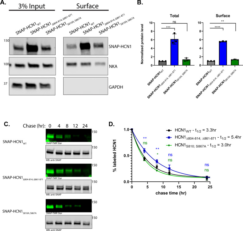
Hyperpolarization activated cyclic nucleotide-gated channel 1 (HCN1) is expressed throughout the nervous system and is critical for regulating neuronal excitability, with mutations being associated with multiple forms of epilepsy. Adaptive modulation of HCN1 has been observed, as has pathogenic dysregulation. While the mechanisms underlying this modulation remain incompletely understood, regulation of HCN1 has been shown to include phosphorylation. A candidate phosphorylation-dependent regulator of HCN1 channels is 14-3-3. We used bioinformatics to identify three potential 14-3-3 binding sites in HCN1. We confirmed that 14-3-3 could pull down HCN1 from multiple tissue sources and used HEK293 cells to detail the interaction. Two sites in the intrinsically disordered C-terminus of HCN1 were necessary and sufficient for a phosphorylation-dependent interaction with 14-3-3. The same region of HCN1 containing the 14-3-3 binding peptides is required for phosphorylation-independent protein degradation. We propose a model in which phosphorylation of mouse S810 and S867 (human S789 and S846) recruits 14-3-3 to inhibit a yet unidentified factor signaling for protein degradation, thus increasing the half-life of HCN1.


Conflict of interest statement

The authors have declared that no competing interests exist.

Figures

Fig 1
Fig 1. The C-terminus of HCN1 contains multiple predicted 14-3-3 binding sites.
Sequence alignment of HCN1 from five vertebrate species. The structured domains of HCN1 are shaded as follows: light gray, HCN domain; dark gray, transmembrane domains; and medium gray, gating ring (C-linker and Cyclic Nucleotide Binding Domain (CNBD)). The RxR ER retention motif is colored in orange and the TRIP8b binding site in cyan. 14-3-3Pred predicted 14-3-3 binding sites are underlined and the three sites that were tested in this study are further shaded in yellow.
Fig 2
Fig 2. 14-3-3 interacts with HCN1.
A) Western blotting for HCN1 and 14-3-3 following 14-3-3 IP or control GFP IP from wild-type and HCN1 KO retina. B) Western blotting for HCN1 following pulldown with recombinant 14-3-3ζ or resin only control from mouse retina, whole brain, and HCN1 expressing HEK293 cells.
Fig 3
Fig 3. Mutating 14-3-3 interaction sites enhances HCN1 protein level in stable HEK293 cell lines.
Activation voltage (A), current density (B), and maximal current density of Ih (C), recorded from HEK293 cell lines expressing mRuby3-HCN1 (black) or mRuby3-HCN1Δ3, 14-3-3 (grey). D) Representative western blot showing total HCN1 expression and surface HCN1 expression isolated by surface biotinylation. Western blotting against the endogenous NKA and GAPDH as membrane and cytosolic protein controls. E) Relative amount of total and surface mRuby3-HCN1Δ3, 14-3-3 compared to mRuby3-HCN1 normalized to NKA. Error bars represent standard error of mean for electrophysiology data (A, B, C) and standard deviation for western blot data (E). T-test p < 0.05 (*); p < 0.01 (**); p<0.001 (***); p < 0.0001 (****).
Fig 4
Fig 4. 14-3-3 interacts with two sites in the distal C-terminus of HCN1.
A) Schematic of the reporter system used in this series of experiments. The C-terminus of HCN1 (residues 598–910) was fused to the transmembrane reporter. Reporters were expressed in HEK293 cells and pulled down with recombinant 14-3-3ζ then analyzed by western blotting for the SNAP-tag. B) Summary of reporters used. Binding is given as efficiency compared to full-length HCN1 C-terminus with ++++ = 100%, +++ = 75%, ++ = 50%, + = 25%, and— = 0% binding efficiency. C, E, G, I) Representative blots and comparisons of normalized results (D, F, H, J) for pulldowns of the reporter-HCNCT constructs as indicated. K) representative pulldown L) comparison of pulldown from cells CT730-910 treated with the broad-spectrum kinase inhibitor staurosporine. For each construct, the p-value or adjusted p-value for comparison to CT598-910 or CT731-910 or untreated control is shown p < 0.05 (*); p < 0.01 (**); p<0.001 (***); p < 0.0001 (****).
Fig 5
Fig 5. 14-3-3 interaction sites regulate HCN1 turnover in a phosphorylation independent manner.
A) Representative western blot showing total SNAP-HCN1 expression and surface SNAP-HCN1 expression isolated by surface biotinylation with western blotting against the endogenous NKA and GAPDH as membrane and cytosolic protein controls. B) Relative total and surface level of SNAP-HCN1Δ804–814; Δ861–871 (blue) and SNAP-HCN1S810A; S867A (green) compared to SNAP-HCN1 (black) normalized to NKA. C) Representative fluorescence imaging of SNAP-HCN1 harvested after 0, 4, 8, 12, and 24 hours of chase. SNAP-TMR Star was used to detect labeled SNAP-HCN1 and anti-SNAP antibody for total SNAP-HCN1. D) Relative amount of labeled SNAP-HCN1 (black), SNAP-HCN1Δ804–814; Δ861–871 (blue), and SNAP-HCN1S810A; S867A (green), normalized to amount at 0 hour chase plotted against chase time to give a decay curve. The adjusted p-value for comparison to SNAP-HCN1 is shown p < 0.05 (*); p < 0.01 (**); p<0.001 (***); p < 0.0001 (****).
Fig 6
Fig 6. Potential Model for HCN1 degradation versus 14-3-3 interaction with HCN1.
A) One potential model that could explain the apparent dual nature of the S810 & S867 centered signals in the HCN1 C-terminus–the serines are required for interaction with 14-3-3 and the flanking sequences around those serines participate in HCN1 degradation by an unknown factor (X). HCN1 phosphorylation at S810 and S867 could bias protein-protein interactions at those sites towards 14-3-3 over the Factor X degradation mediator. B) HCN1 protein level is established by the balance between destabilizing forces, such as the strong degradation signal, and stabilizing forces. The role of 14-3-3 interaction with HCN1 remains unclear but it could act in combination with other proteins, such as the well-established neuron specific HCN1 binding partner TRIP8b to provide a modularized strong protection signal to balance the effects of the degradation signal.

References

    1. Biel M, Wahl-Schott C, Michalakis S, Zong X. Hyperpolarization-activated cation channels: from genes to function. Physiological reviews. 2009;89(3):847–85. doi: 10.1152/physrev.00029.2008 . - DOI - PubMed
    1. He C, Chen F, Li B, Hu Z. Neurophysiology of HCN channels: from cellular functions to multiple regulations. Prog Neurobiol. 2014;112:1–23. Epub 2013/11/05. doi: 10.1016/j.pneurobio.2013.10.001 . - DOI - PubMed
    1. Shah MM. Cortical HCN channels: function, trafficking and plasticity. J Physiol. 2014;592(13):2711–9. Epub 2014/04/24. doi: 10.1113/jphysiol.2013.270058 ; PubMed Central PMCID: PMC4104471. - DOI - PMC - PubMed
    1. Bonzanni M, DiFrancesco JC, Milanesi R, Campostrini G, Castellotti B, Bucchi A, et al.. A novel de novo HCN1 loss-of-function mutation in genetic generalized epilepsy causing increased neuronal excitability. Neurobiol Dis. 2018;118:55–63. Epub 2018/06/25. doi: 10.1016/j.nbd.2018.06.012 . - DOI - PubMed
    1. Marini C, Porro A, Rastetter A, Dalle C, Rivolta I, Bauer D, et al.. HCN1 mutation spectrum: from neonatal epileptic encephalopathy to benign generalized epilepsy and beyond. Brain. 2018;141(11):3160–78. Epub 2018/10/24. doi: 10.1093/brain/awy263 . - DOI - PubMed

Publication types

MeSH terms

Substances