. 2022 Jun 27;62(12):3023-3033.
doi: 10.1021/acs.jcim.2c00348. Epub 2022 Jun 9.

Alchemical Free Energy Calculations to Investigate Protein-Protein Interactions: the Case of the CDC42/PAK1 Complex

Affiliations

Alchemical Free Energy Calculations to Investigate Protein-Protein Interactions: the Case of the CDC42/PAK1 Complex

Maria Antonietta La Serra et al. J Chem Inf Model. .

Abstract

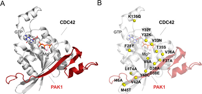
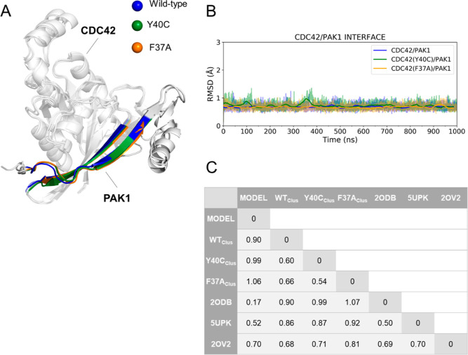
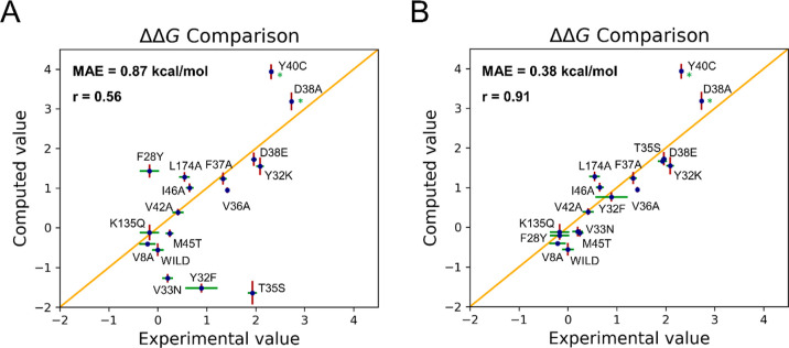
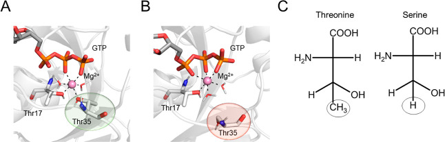
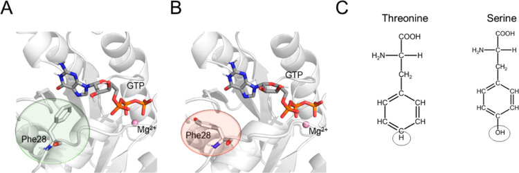
Here, we show that alchemical free energy calculations can quantitatively compute the effect of mutations at the protein-protein interface. As a test case, we have used the protein complex formed by the small Rho-GTPase CDC42 and its downstream effector PAK1, a serine/threonine kinase. Notably, the CDC42/PAK1 complex offers a wealth of structural, mutagenesis, and binding affinity data because of its central role in cellular signaling and cancer progression. In this context, we have considered 16 mutations in the CDC42/PAK1 complex and obtained excellent agreement between computed and experimental data on binding affinity. Importantly, we also show that a careful analysis of the side-chain conformations in the mutated amino acids can considerably improve the computed estimates, solving issues related to sampling limitations. Overall, this study demonstrates that alchemical free energy calculations can conveniently be integrated into the design of experimental mutagenesis studies.


Conflict of interest statement

The authors declare no competing financial interest.

Figures

Figure 1
Figure 1
Structural representation of the CDC42/PAK1 model. (A) CDC42/PAK1 complex is reported (see Methods section). CDC42 is represented as a white cartoon, while PAK1 is shown in red. The GTP nucleotide and the Mg2+ ion are in sticks and balls, respectively. (B) Analyzed single-point mutations are represented as yellow balls on the CDC42/PAK1 complex structure.
Figure 2
Figure 2
RMSD analysis of the interface of the CDC42/PAK1 systems. (A) Structural alignment of representative conformations from MD simulations. The protein structures are represented as a white cartoon, while the GTP nucleotide and Mg2+ ion are illustrated as sticks and balls, respectively. Interface residues on both CDC42 and PAK1 are highlighted as blue, green, and orange for the wt, Y40C, and F37A systems, respectively. (B) Time-series RMSD descriptors for CDC42 wt (blue), Y40C (green), and F37A (orange) variants are reported. (C) RMSD (in Å) of interface residues between different structures, including MD representative structures, the initial model, and experimental structures.
Figure 3
Figure 3
(A) Initial ΔΔGb (in kcal/mol) computed using the alchemical transformations and plotted against the experimental values. (B) Scatter plot obtained after improving the ΔΔGb estimates for T35S, F28Y, Y32F, and V33N (see text for details). In both A and B, the examined single-point mutations are reported together with their computed (red) and experimental (green) error bars. The asterisk (*) marks mutations for which the experimental error was not reported.
Figure 4
Figure 4
T35S CDC42 variant. (A) Interaction between the catalytic Mg2+ and the hydroxyl group of Thr35 as observed in the equilibrium MD simulations of the wt system; (B) after the initial alchemical transformation of Thr25 into Ser, the coordination sphere of Mg2+ was disrupted. The protein is represented as a white cartoon, the GTP nucleotide and the residues coordinating Mg2+ as sticks, and the Mg2+ ion as a ball. (C) Revised atom mapping used to improve the ΔΔGb estimate retrieved from alchemical transformation. In this case, the circled atoms are those considered unique for the transformation.
Figure 5
Figure 5
F28Y CDC42 variant. (A) Interaction between GTP and the aromatic ring of Phe28 as observed in the equilibrium MD simulations of the wt system; (B) after the initial alchemical transformation of Phe28 into Tyr, the aromatic ring was no longer in contact with GTP. The protein is represented as a white cartoon, the GTP nucleotide and the residues involved in the single-point mutation as sticks, and the Mg2+ ion as a ball. (C) Revised atom mapping used to improve the ΔΔGb estimate retrieved from the alchemical transformation. In this case, the circled atoms are those considered unique for the transformation.
Figure 6
Figure 6
Conformational analysis of Y32 and V33. (A) Distribution of the distance between Tyr32 and the γ-phosphate of GTP. The green and red color codes indicate the most and the least populated Tyr32 conformations, respectively. In the upper right panel, representative conformations of the side chain of Tyr32 observed during the equilibrium MD simulations of the wt system are reported. CDC42 is represented as a white cartoon, Tyr32 and the GTP nucleotide in sticks, and the Mg2+ ion as a ball. (B) Distribution of the N–CA–CB–CG1 V33 dihedral angle. In the upper right panel, a representative conformation of the side chain of Val33 as observed during the equilibrium MD simulations of the wt system is reported. CDC42 is represented as a white cartoon, while Val33 is shown as sticks.

References

    1. Pawson T.; Nash P. Protein–Protein Interactions Define Specificity in Signal Transduction. Genes Dev. 2000, 14, 1027–1047. 10.1101/GAD.14.9.1027. - DOI - PubMed
    1. Keskin O.; Gursoy A.; Ma B.; Nussinov R. Principles of Protein–Protein Interactions: What Are the Preferred Ways for Proteins to Interact?. Chem. Rev. 2008, 108, 1225–1244. 10.1021/cr040409x. - DOI - PubMed
    1. Ivarsson Y.; Jemth P. Affinity and Specificity of Motif-Based Protein–Protein Interactions. Curr. Opin. Struct. Biol. 2019, 54, 26–33. 10.1016/J.SBI.2018.09.009. - DOI - PubMed
    1. Borgia A.; Borgia M. B.; Bugge K.; Kissling V. M.; Heidarsson P. O.; Fernandes C. B.; Sottini A.; Soranno A.; Buholzer K. J.; Nettels D.; Kragelund B. B.; Best R. B.; Schuler B. Extreme Disorder in an Ultrahigh-Affinity Protein Complex. Nature 2018, 555, 61–66. 10.1038/nature25762. - DOI - PMC - PubMed
    1. Lu H.; Zhou Q.; He J.; Jiang Z.; Peng C.; Tong R.; Shi J. Recent Advances in the Development of Protein–Protein Interactions Modulators: Mechanisms and Clinical Trials. Signal Transduction Targeted Ther. 2020, 5, 1–23. 10.1038/s41392-020-00315-3. - DOI - PMC - PubMed

Publication types