. 2022 Aug 9;6(15):4360-4372.
doi: 10.1182/bloodadvances.2022007975.

In vivo HSC transduction in rhesus macaques with an HDAd5/3+ vector targeting desmoglein 2 and transiently overexpressing cxcr4

Affiliations

In vivo HSC transduction in rhesus macaques with an HDAd5/3+ vector targeting desmoglein 2 and transiently overexpressing cxcr4

Hongjie Wang et al. Blood Adv. .

Abstract

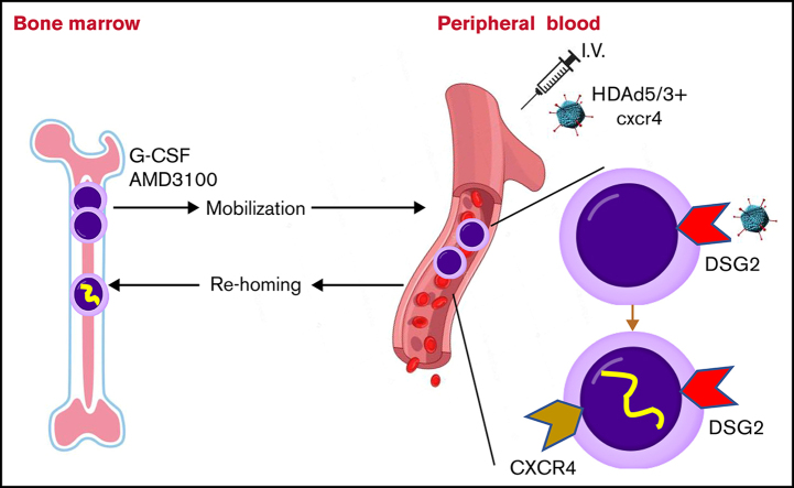
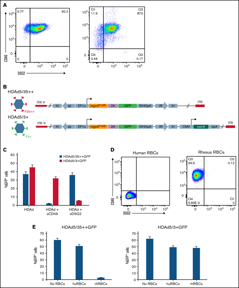
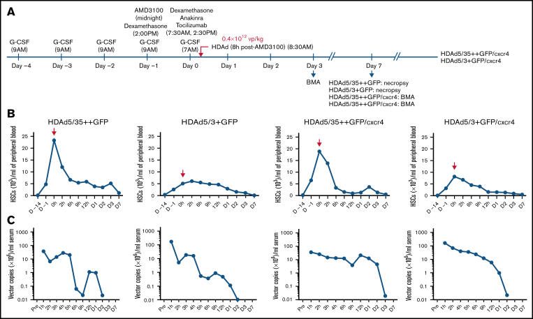
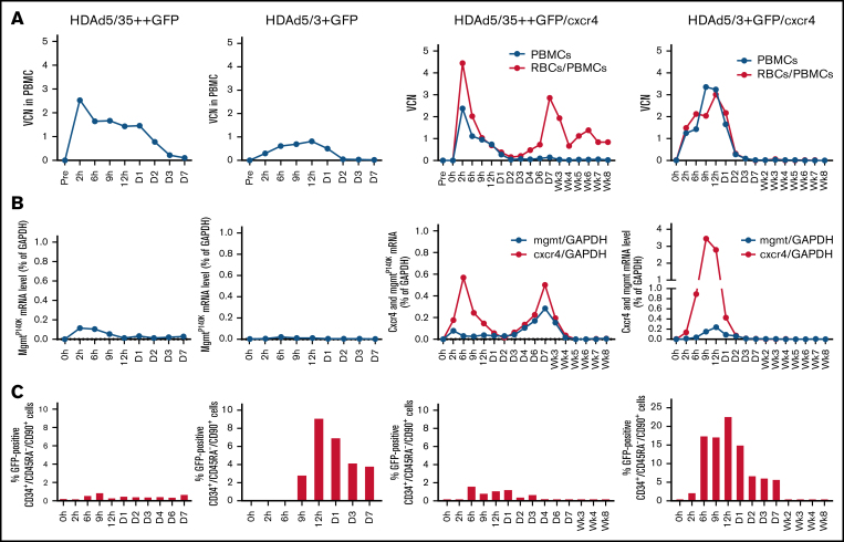
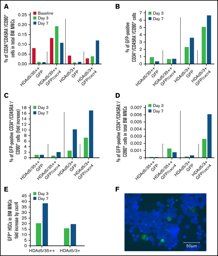
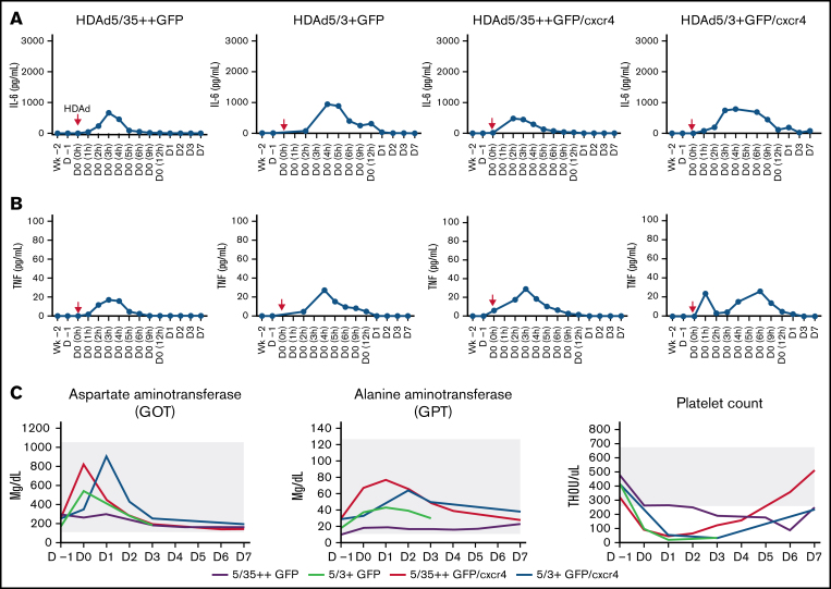
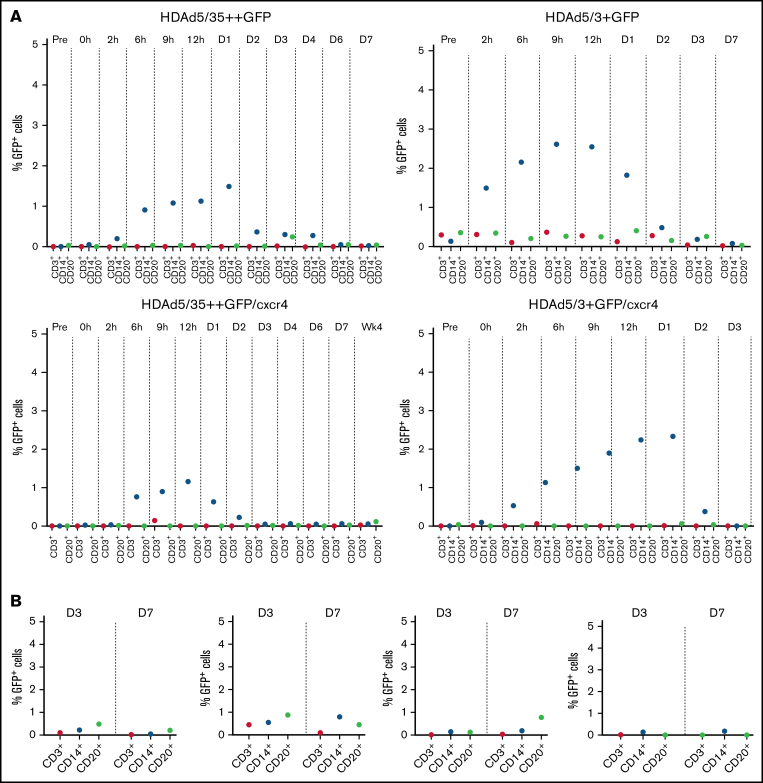
We developed a new in vivo hematopoietic stem cell (HSC) gene therapy approach that involves only IV injections and does not require myeloablation/conditioning and HSC transplantation. In this approach, HSCs are mobilized from the bone marrow into the peripheral bloodstream and transduced with IV injected helper-dependent adenovirus (HDAd) vectors. A fraction of transduced HSCs returns to the bone marrow and persists there long term. Here, we report desmoglein 2 (DSG2) as a new receptor that can be used for in vivo HSC transduction. HDAd5/3+ vectors were developed that use DSG2 as a high-affinity attachment receptor, and in vivo HSC transduction and safety after IV injection of an HDAd5/3+ vector expressing green fluorescent protein (GFP) in granulocyte colony-stimulating factor/AMD3100 (plerixafor)-mobilized rhesus macaques were studied. Unlike previously used CD46-targeting HDAd5/35++ vectors, HDAd5/3+ virions were not sequestered by rhesus erythrocytes and therefore mediated ∼10-fold higher GFP marking rates in primitive HSCs (CD34+/CD45RA-/CD90+ cells) in the bone marrow at day 7 after vector injection. To further increase the return of in vivo transduced, mobilized HSCs to the bone marrow, we transiently expressed cxcr4 in mobilized HSCs from the HDAd5/3+ vector. In vivo transduction with an HDAd5/3+GFP/cxcr4 vector at a low dose of 0.4 × 10¹² viral particles/kg resulted in up to 7% of GFP-positive CD34+/CD45RA-/CD90+ cells in the bone marrow. This transduction rate is a solid basis for in vivo base or prime editing in combination with natural or drug-induced expansion of edited HSCs. Furthermore, our study provides new insights into HSC biology and trafficking after mobilization in nonhuman primates.


Figures

None
Graphical abstract
Figure 1
Figure 1
Analysis of DSG2 expression on HSCs. (A) Flow cytometry for CD46 and DSG2 on human CD34+ cells from 2 different healthy G-CSF–mobilized donors. (B) Schematic of HDAd vector capsids and genomes. The chimeric capsids contain either affinity-enhanced Ad35 or Ad3 fiber knob domains. Both vector genomes contain an EF1α-mgmtp140k/GFP expression cassette used before. Both proteins are linked via a self-cleaving picornavirus 2A peptide. One set of vectors has an additional CMV promoter–human cxcr4 expression cassette. The 4 vectors used in this study are HDAd5/35++GFP, HDAd5/3+GFP, HDAd5/35++GFP/cxcr4, and HDAd5/3+GFP/cxcr4. (C) In vitro CD34+ cell transduction studies. Cells were infected with HDAd5/35++-GFP and HDAd5/3+-GFP virus at a multiplicity of infection of 2000 vp/cell with and without pre-incubation with recombinant soluble CD46 or DSG2 (10 μg/mL) for 1 hour. GFP expression was analyzed 24 hours' postinfection. (D) CD46 and DSG2 flow cytometry on human and rhesus peripheral RBCs stained with antibodies that recognize the receptors in both species (anti-human CD46 mAb clone M177 from Santa Cruz Biotechnology [Dallas, TX] and anti-human DSG2 polyclonal antibody AF947 from R&D Systems [Minneapolis, MN]). (E) Transduction of 293 cells with HDAd5/35++GFP and HDAd5/3+GFP in the absence and presence of human (huRBCs) and rhesus (rhRBCs) RBCs (details are provided in the Materials and methods). Shown is the percentage of GFP-positive cells measured 2 days after transduction.
Figure 2
Figure 2
In vivo HSC transduction studies in rhesus macaques with HDAd5/35++GFP, HDAd5/3+GFP, HDAd5/35++GFP/cxcr4, and HDAd5/3+GFP/cxcr4. (A) Schematic of the experiment showing the injection time of the mobilization drugs (G-CSF and AMD3100) and cytokine prophylaxis drugs (dexamethasone, anakinra, and tocilizumab), as well as the injection time of the HDAd vector at a dose of 0.4 × 10¹² vp/kg. The time point of HDAd injection is taken as “day 0, 0 hours.” HDAd5/35++GFP– and HDAd5/3+GFP–injected animals were monitored for 7 days. HDAd5/35++GFP/cxcr4– and HDAd5/3+GFP/cxcr4–injected animals were observed for 8 weeks to capture late effects of cxcr4 expression. Bone marrow aspirates (BMAs) were performed at days 3, 7, and (for HDAd5/35++GFP/cxcr4 and HDAd5/3+GFP/cxcr4) at weeks 4 and 8. Blood samples were taken at 0 hours (before HDAd injection) and 2, 6, 9, and 12 hours on day 0; on days 1, 2, 3, and 7; and then weekly. (B) HSC mobilization. Shown are numbers of primitive (CD34+/CD45RA/CD90+) HSCs in peripheral blood. HDAd vectors were IV injected at day 0, 0 hours. Notably, there was no correlation between the weight of the animals and the efficacy of mobilization. The weights were 9.70 kg (HDAd5/35++GFP), 6.64 kg (HDAd5/3+GFP), 7.13 kg (HDAd5/35++GFP/cxcr4), and 7.19 kg (HDAd5/3+GFP/cxcr4). (C) Vector clearance from blood. HDAd vector genome copies in serum samples were measured by using quantitative polymerase chain reaction.
Figure 3
Figure 3
Analysis of transduction in peripheral blood cells. (A) VCN per cell in PBMCs (black lines) and total blood cells (RBCs+PBMCs, red lines, measured only for HDAd5/35++GFP/cxcr4– and HDAd5/3+GFP/cxcr4–injected animals). (B) Analysis of transgene mRNA levels relative to mRNA levels of the housekeeping gene GAPDH. Human mgmtP140K mRNA data are shown by black lines; human cxcr4 mRNA levels are shown by red lines. Note that the scale of the y-axis for the HDAd5/3+GFP/cxcr4–injected animal is different. (C) Percentage of GFP+ HSCs in peripheral blood white cells. The scale of the y-axis for the HDAd5/3+GFP/cxcr4–injected animal is different.
Figure 4
Figure 4
GFP expression in bone marrow (BM) HSCs and MNCs. (A) Return of mobilized HSCs to the BM. Shown is the percentage of primitive (CD34+/CD45RA/CD90+) HSCs in total BM MNCs measured in BM samples at days 3 and 7. (“Day 0” is the day of HDAd injection.) (B) In vivo transduced primitive HSCs that returned to the BM. Shown is the percentage of GFP-positive CD34+/CD45RA/CD90+ HSCs in BM aspirates collected at days 3 and 7. (C) Shown are the data from panel B as fold increase, taking transduction with the HDAd5/35++GFP vector as “1.” (D) Percentage of GFP-positive CD34+/CD45RA/CD90+ HSCs relative to total BM MNCs. (E) Data extracted from panel D showing the effect of cxcr4 expression on the GFP+ HSCs in the BM (comparison of HDAd5/35++GFP vs HDAd5/35++GFP/cxcr4 and HDAd5/3+GFP vs HDAd5/3+GFP/cxcr4). (F) Representative GFP immunofluorescence on a BM CD34+ cytospin of the HDAd5/3+GFP/cxcr4–injected animal. CD34+ cells were isolated from a BM aspirate (after RBC lysis) by human CD34 magnetic-activated cell sorting, spun on a glass slide, and then stained with a GFP–fluorescein isothiocyanate antibody. GFP signals are green. Nuclei were stained with 4′,6-diamidino-2-phenylindole and appear in blue. Scale bar = 50 μm.
Figure 5
Figure 5
Safety of IV HDAd injection into mobilized rhesus macaques. (A) Serum IL-6 levels measured by cytometric bead array. HDAd injections (day 0, 0 hours) are indicated by red arrows. (B) Serum TNFα levels. (C) Selected hematologic parameters. The normal range is indicated by the gray shading. THOU, thousands.
Figure 6
Figure 6
Percentage of GFP-positive cells in lineage–positive cells. Percentage of GFP+ CD3+, CD14+, and CD20+ lineage cells in PBMCs (A) and bone marrow MNCs (B).

References

    1. Wang H, Georgakopoulou A, Li C, et al. Curative in vivo hematopoietic stem cell gene therapy of murine thalassemia using large regulatory elements. JCI Insight. 2020;5(16):139538. - PMC - PubMed
    1. Li C, Wang H, Georgakopoulou A, Gil S, Yannaki E, Lieber A. In vivo HSC gene therapy using a bi-modular HDAd5/35++ vector cures sickle cell disease in a mouse model. Mol Ther. 2021;29(2):822–837. - PMC - PubMed
    1. Li C, Georgakopoulou A, Mishra A, et al. In vivo HSPC gene therapy with base editors allows for efficient reactivation of fetal γ-globin in β-YAC mice. Blood Adv. 2021;5(4):1122–1135. - PMC - PubMed
    1. Li C, Psatha N, Sova P, et al. Reactivation of γ-globin in adult β-YAC mice after ex vivo and in vivo hematopoietic stem cell genome editing. Blood. 2018;131(26):2915–2928. - PMC - PubMed
    1. Wang H, Georgakopoulou A, Psatha N, et al. In vivo hematopoietic stem cell gene therapy ameliorates murine thalassemia intermedia. J Clin Invest. 2019;129(2):598–615. - PMC - PubMed

Publication types

MeSH terms