Review
. 2022 Jun 14;39(11):110969.
doi: 10.1016/j.celrep.2022.110969. Epub 2022 May 30.

Emerging viruses: Cross-species transmission of coronaviruses, filoviruses, henipaviruses, and rotaviruses from bats

Affiliations
Review

Emerging viruses: Cross-species transmission of coronaviruses, filoviruses, henipaviruses, and rotaviruses from bats

Jin Tian et al. Cell Rep. .

Abstract

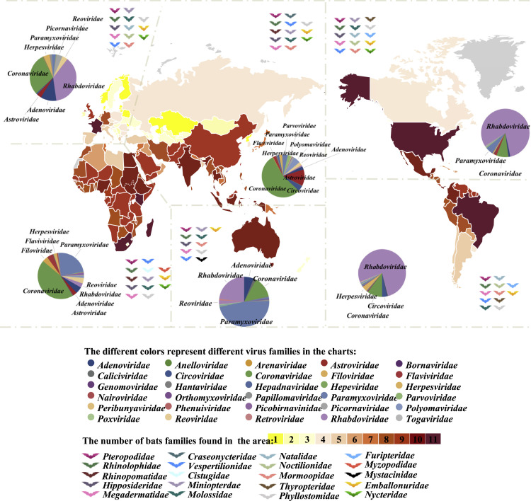
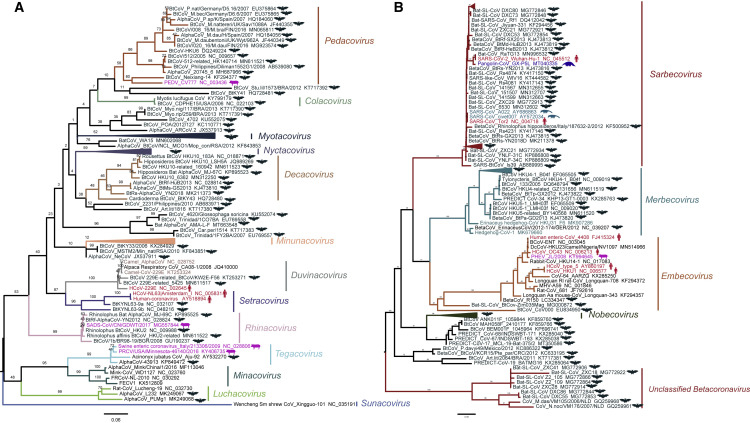
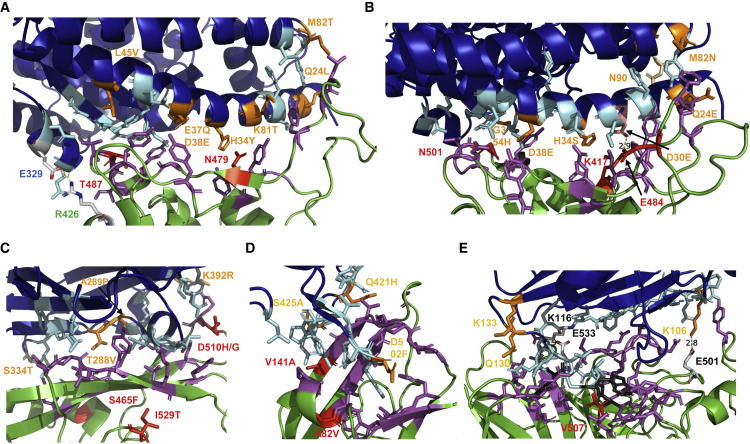
Emerging infectious diseases, especially if caused by bat-borne viruses, significantly affect public health and the global economy. There is an urgent need to understand the mechanism of interspecies transmission, particularly to humans. Viral genetics; host factors, including polymorphisms in the receptors; and ecological, environmental, and population dynamics are major parameters to consider. Here, we describe the taxonomy, geographic distribution, and unique traits of bats associated with their importance as virus reservoirs. Then, we summarize the origin, intermediate hosts, and the current understanding of interspecies transmission of Middle East respiratory syndrome coronavirus (MERS-CoV), severe acute respiratory syndrome coronavirus (SARS-CoV), SARS-CoV-2, Nipah, Hendra, Ebola, Marburg virus, and rotaviruses. Finally, the molecular interactions of viral surface proteins with host cell receptors are examined, and a comparison of these interactions in humans, intermediate hosts, and bats is conducted. This uncovers adaptive mutations in virus spike protein that facilitate cross-species transmission and risk factors associated with the emergence of novel viruses from bats.

Keywords: CP: Microbiology; RNA virus; bat; interspecies transmission.


Conflict of interest statement

Declaration of interests The authors declare no competing interests.

Figures

Figure 1
Figure 1
Global distribution of bats and their viruses The bat families present in each continent are symbolized by the color of the bat pictogram. The number of different bat families identified in each country is represented by the color range (from yellow to red) as indicated. The pie charts show the percentage of bat-associated viruses from a certain family identified in the respective continents according to publicly available sequence data from a database of bat-associated viruses (http://www.mgc.ac.cn/DBatVir/) as of July 16, 2020, and GenBank data from July 16, 2020 to August 25, 2020.
Figure 2
Figure 2
Genetic evolution of coronaviruses in Alphacoronavirus and Betacoronavirus (A and B) Maximum likelihood tree of Alphacoronavirus (A) and Betacoronavirus (B) reconstructed by RAxML based on the polymerase (RdRP) gene, using the PROTCATLG substitution model and 1,000 bootstrap value. Representative isolate sequences used in the present study were downloaded from GenBank and the RdRP gene was intercepted, and the corresponding GenBank accession numbers were provided in the tree. The hosts of the viruses are indicated by different colors (bat, black; human, dark red; pig, magenta; pangolin, blue; and civet, dark green) and by pictograms in the same color. Trees were rooted on midpoint, and branches of the tree labeled in different colors indicate the indicated subgenera.
Figure 3
Figure 3
Interaction of SARS-CoV, SARS-CoV-2, MERS-CoV, EBOV, and NiV with their receptors (A) Interaction surface between ACE2 and S of SARS-CoV: ACE2 is colored blue, S in green. The contacting amino acids are shown as cyan (ACE2) or magenta (S) sticks. The seven amino acids substituted in civet ACE2 are labeled in orange. In ACE2 of mice, M82 is substituted to N, thereby creating an N-glycosylation site. The two amino acid substitutions during adaptation of SARS-CoV from civets to humans (N479) and from humans to humans (T487) are shown in red. Glu329 in S and Arg426 in ACE2 form a salt bridge at the periphery of the interaction surface. This figure was generated with PyMol 2.1.1. using the pdb file 2AJF. The contacting amino acids shown here are from Lan et al. (2020), which differ in four peripheral residues from the first publication Li et al. (2005a). (B) Interaction surface between ACE2 and S of SARS-CoV-2: ACE2 is colored blue, S in green. The contacting amino acids are shown as cyan (ACE2) or magenta (S) sticks. The seven amino acids substituted in pangolin ACE2 are labeled in orange. Lys417 in S and Glu30 in ACE2 form a salt bridge in the middle of the interaction surface. Lys417 (labeled red) is substituted by a Thr in variant P1 and by an Asn in variant B 1.351. Highlighted as a red stick is also N501, which is substituted by Tyr in variants B1.1.1, B1.351. and P1. The figure was generated with PyMol 2.1.1. using the pdb file 6M0J. (C) Interaction surface between human DPP4 and S of MERS-CoV: DPP4 is colored blue, S in green. The contacting amino acids are shown as cyan (DPP4) or magenta (S) sticks. The four amino acids substituted in camel DPP4 are labeled in orange. Residue 334 is not directly contacting the spike but is an N-glycosylation site in mice DPP4, which needs to be removed to make mice susceptible to MERS-CoV infection. The two amino acid substitutions (S465F, D510H) during adaptation of S to a suboptimal bat-associated virus receptor and during MERS outbreak in South Korea (I529T, D510G) are shown in red. The figure was generated with PyMol 2.1.1. using the pdb file 4KRO. (D) Interaction surface between human NPC1 and GP of EBOV. NPC1 is colored blue, GP in green. The contacting amino acids are shown as cyan (NPC1) or magenta (GP) sticks. The three amino acids substituted in pig NPC1 are labeled in orange. Amino acid substitutions restoring EBOV binding to refractory receptors (V141A) and during the large Ebola epidemic (A82V) are labeled red. The figure was generated with PyMol 2.1.1. using the pdb file 5F1B. (E) Interaction surface between human ephrin-b2 and G of Nipah virus. Ephrin-B2 is colored blue, G in green. Most of the contacting amino acids are shown as sticks. Three hydrophobic amino acids (F120, L124, W125) in the loop of ephrin-B2 essential for binding are colored in gray. The two salt bridges (K106 with E501, K116 with E533) are indicated. The only amino acid substitution between G of Nipah and Hendra virus is shown in red. Receptor amino acids near the binding site variable between species (K106, Q130, K133) are labeled in orange. The figure was generated with PyMol 2.1.1. using the pdb file 2VSM.

References

    1. Adney D.R., van Doremalen N., Brown V.R., Bushmaker T., Scott D., de Wit E., Bowen R.A., Munster V.J. Replication and shedding of MERS-CoV in upper respiratory tract of inoculated dromedary camels. Emerg. Infect. Dis. 2014;20:1999–2005. doi: 10.3201/eid2012.141280. - DOI - PMC - PubMed
    1. Amendola A., Bianchi S., Gori M., Colzani D., Canuti M., Borghi E., Raviglione M.C., Zuccotti G.V., Tanzi E. Evidence of SARS-CoV-2 RNA in an oropharyngeal swab specimen, milan, Italy, early December 2019. Emerg. Infect. Dis. 2021;27:648–650. doi: 10.3201/eid2702.204632. - DOI - PMC - PubMed
    1. Annan A., Baldwin H.J., Corman V.M., Klose S.M., Owusu M., Nkrumah E.E., Badu E.K., Anti P., Agbenyega O., Meyer B., et al. Human betacoronavirus 2c EMC/2012-related viruses in bats, Ghana and Europe. Emerg. Infect. Dis. 2013;19:456–459. doi: 10.3201/eid1903.121503. - DOI - PMC - PubMed
    1. Anthony S.J., Epstein J.H., Murray K.A., Navarrete-Macias I., Zambrana-Torrelio C.M., Solovyov A., Ojeda-Flores R., Arrigo N.C., Islam A., Ali Khan S., et al. A strategy to estimate unknown viral diversity in mammals. mBio. 2013;4:e00598-13. doi: 10.1128/mbio.00598-13. - DOI - PMC - PubMed
    1. Anthony S.J., Gilardi K., Menachery V.D., Goldstein T., Ssebide B., Mbabazi R., Navarrete-Macias I., Liang E., Wells H., Hicks A., et al. Further evidence for bats as the evolutionary source of middle east respiratory syndrome coronavirus. mBio. 2017;8:e00373-17. - PMC - PubMed

Publication types