. 2022 Aug;28(8):1144-1155.
doi: 10.1261/rna.079173.122. Epub 2022 Jun 9.

RNase H-based analysis of synthetic mRNA 5' cap incorporation

Affiliations

RNase H-based analysis of synthetic mRNA 5' cap incorporation

S Hong Chan et al. RNA. 2022 Aug.

Abstract

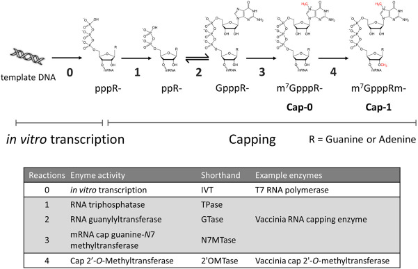
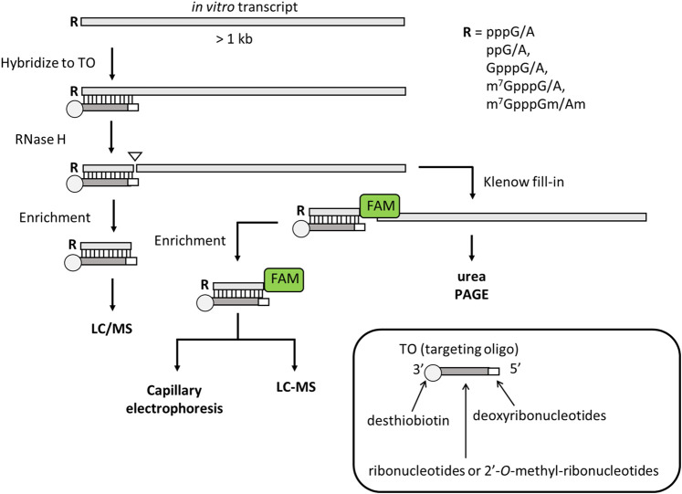
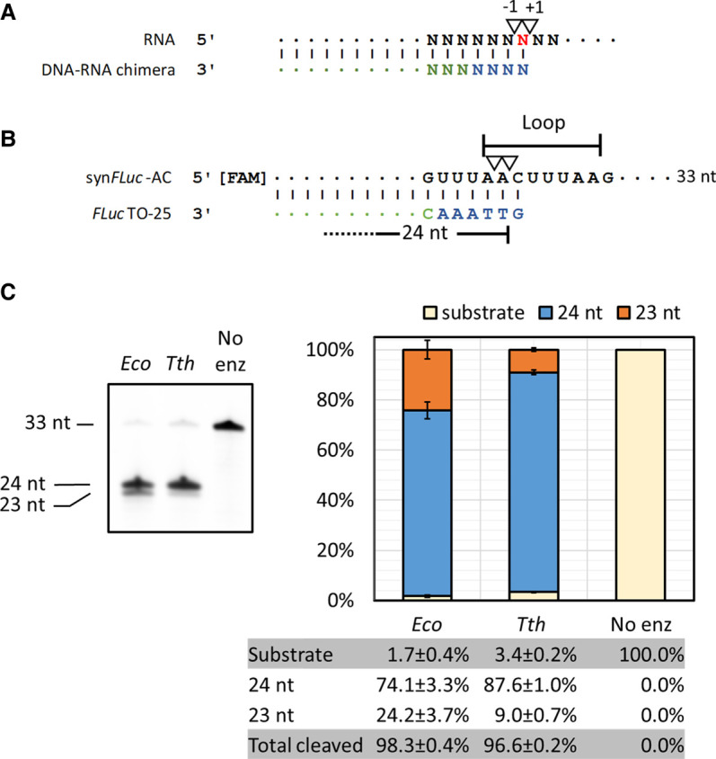
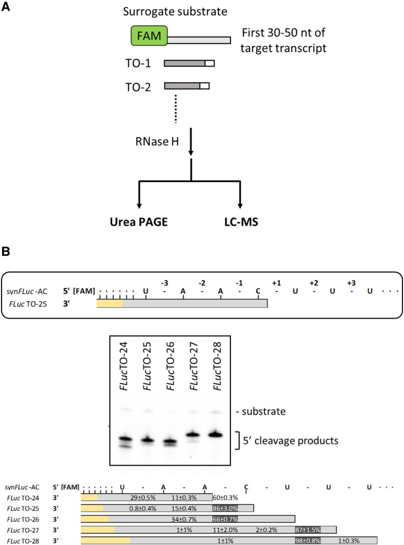
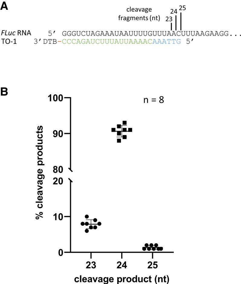
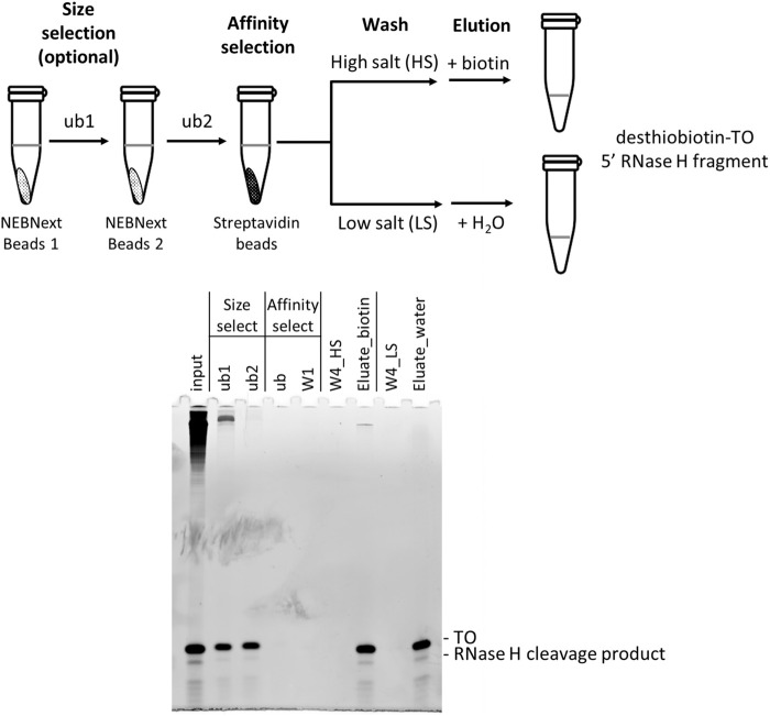
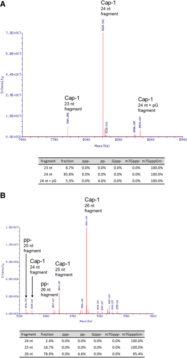
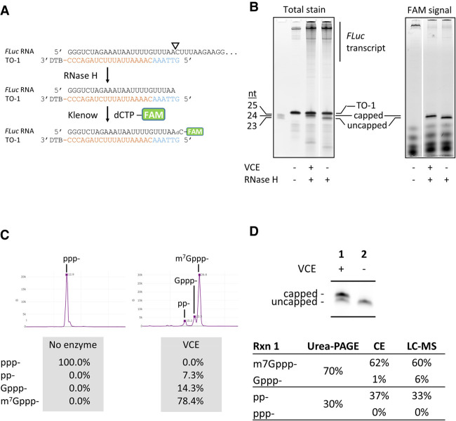
Advances in mRNA synthesis and lipid nanoparticles technologies have helped make mRNA therapeutics and vaccines a reality. The 5' cap structure is a crucial modification required to functionalize synthetic mRNA for efficient protein translation in vivo and evasion of cellular innate immune responses. The extent of 5' cap incorporation is one of the critical quality attributes in mRNA manufacturing. RNA cap analysis involves multiple steps: generation of predefined short fragments from the 5' end of the kilobase-long synthetic mRNA molecules using RNase H, a ribozyme or a DNAzyme, enrichment of the 5' cleavage products, and LC-MS intact mass analysis. In this paper, we describe (1) a framework to design site-specific RNA cleavage using RNase H; (2) a method to fluorescently label the RNase H cleavage fragments for more accessible readout methods such as gel electrophoresis or high-throughput capillary electrophoresis; (3) a simplified method for post-RNase H purification using desthiobiotinylated oligonucleotides and streptavidin magnetic beads followed by elution using water. By providing a design framework for RNase H-based RNA 5' cap analysis using less resource-intensive analytical methods, we hope to make RNA cap analysis more accessible to the scientific community.

Keywords: 5′ cap; LC-MS; RNA cap analysis; RNA capping; RNase H; synthetic mRNA.


Figures

FIGURE 1.
FIGURE 1.
Enzymatic reactions involved in RNA 5′ capping. RNA transcripts generated by in vitro transcription (usually done using T7 RNA polymerase or its variants; reaction 0) contains a triphosphate group at the 5′ end (pppR-). The 5′ triphosphate group can be converted to the Cap-1 structure (m7GpppRm-) through four enzymatic reactions carried out by RNA capping enzymes such as vaccinia RNA capping enzyme (reactions 1 through 3) in conjunction with vaccinia cap 2′-O-methyltransferase (reaction 4).
FIGURE 2.
FIGURE 2.
A general scheme of RNase H-based RNA cap analysis. A DNA–RNA or DNA-2′-O-methyl-ribonucleotide chimera is designed to be complementary to part of the 5′ end of the target RNA molecule such that the chimera stays annealed to the cleavage fragment after RNase H cleavage. The chimera (called targeting oligo or TO in this paper) contains a 3′-desthiobiotin group. After denaturation and annealing, RNase H cleaves at a predefined site within the RNA-TO duplex and generates a one-base recessive end at the 3′ end of the cleaved RNA. Because RNase H cleavage results in a 3′ hydroxyl group (24), this recessive 3′ end can be filled in with a fluorescently labeled deoxynucleotide using the Klenow fragment of DNA polymerase I. The fluorescently labeled 5′ cleavage fragment can be analyzed by denaturing PAGE directly without enrichment. The 5′ duplex cleavage fragment can be enriched using streptavidin magnetic beads. The enriched RNase H cleavage products can be analyzed by LC-MS or capillary electrophoresis (if filled in with a fluorescent deoxynucleotide).
FIGURE 3.
FIGURE 3.
Directed RNA cleavage with RNase H. (A) Previously reported RNase H cleavage sites (17–19). (B) Synthetic RNA oligonucleotide containing the first 33 nt of an artificial FLuc transcript (synFLuc-AC) and targeting oligos FLucTO-25 designed based on A. Inverted triangles represent RNase H cleavage sites reported in the literature (A) and in this study (B). Deoxynucleotides in TOs are colored in blue; ribonucleotides are colored in green. The predicted loop region of the synthetic RNA oligonucleotides based on an RNA folding algorithm (RNAFold, University of Vienna) is indicated. (C) RNA cleavage using E. coli or Thermus thermophilus RNase H at 37°C. The cleavage efficiency of both enzymes was similar but Tth RNase H generates more uniform cuts than the E. coli enzyme.
FIGURE 4.
FIGURE 4.
Selection of targeting oligos for uniform RNase H cleavage. (A) In general, a surrogate RNA oligonucleotide containing the first 30–50 nt of the target transcript is chemically synthesized with a 5′ FAM group. A series of targeting oligos (TOs) are designed and chemically synthesized. The TOs used in the selection exercise did not require a desthiobiotin group, because unlike the long 3′ cleavage products of in vitro transcripts, the 3′ cleavage products of the surrogate RNA were short and did not interfere with LC-MS analysis. After RNase H cleavage, the fluorescently labeled 5′ cleavage fragments can be analyzed by urea PAGE and LC-MS intact mass analysis. (B) For consistency, the phosphodiester bonds are numbered around the nucleotide hybridized to the 5′ deoxynucleotide of the TO (top panel; a cytidine in this case). Urea PAGE showed that RNase H cleavage is most efficient with FLucTO-25, FLucTO-26, and FLucTO-27. Multiple cleavage products were observed with FLucTO-24, FLucTO-25, and FLucTO-26 (middle panel). LC-MS intact mass analysis of the cleavage products is shown in the lower panel. In the schematics, phosphodiester bonds are shown as “-”. Deoxynucleotides in the TOs are shown as gray bars; ribonucleotides are shown as yellow bars. Numbers represent frequency of cleavage detected at corresponding phosphodiester bonds obtained in triplicated experiments.
FIGURE 5.
FIGURE 5.
Uniform RNase H cleavage with designed targeting oligos. (A) 5′ sequence of a 1.7 kb in vitro FLuc transcript containing an artificial 5′ UTR and the corresponding targeting oligo TO-1. The targeting oligo contains six deoxynucleotides (blue) at the 5′ end followed by 19 ribonucleotides (green) and a desthiobiotin (DTB) group at the 3′ end. The size of the RNase H cleavage products is shown. (B) Frequency of cleavage events expressed as percentage of detected cleavage product using LC-MS. Median of cleavage frequencies was 8%, 91%, and 1% at (A|A), (A|C), and (C|U), respectively.
FIGURE 6.
FIGURE 6.
Enrichment of RNase H cleavage products by size and affinity selection. The RNase H cleavage product (input) was first size-selected by two rounds of NEBNext magnetic beads. The clarified unbound fraction of the second round of size selection (ub2) was then added directly to streptavidin magnetic beads for affinity selection. After the first wash using a standard wash solution containing 1 M NaCl (W1), the resuspended bead slurry was divided into two fractions. One fraction was washed three more times using the standard wash buffer (W4_HS) and eluted using biotin (Eluate_biotin). The other fraction of the slurry was washed three more times using a low NaCl wash solution (W4_LS) followed by elution using the same volume of water (Eluate_water). Similar amounts of RNase H cleavage product and TO were eluted (also see Supplemental Fig. 4).
FIGURE 7.
FIGURE 7.
Deconvoluted mass spectrums of capping analysis. An enzymatically capped Cap-1 CLuc (A) or FLuc transcript (B) was processed with RNase H and the single-step affinity enrichment and analyzed by LC-MS as described in the main text. The RNase H cleavage products with relevant 5′ groups were identified by their distinct deconvoluted mass values. The area under the identified mass peaks was used to calculate the relative percentage of each species in the sample. A mass corresponded to 24 nt + pG in the Cap-1 form was detected in the FLuc transcript. The addition of a pG was probably the result of T7 polymerase slippage at the 5′ end of the transcript, which is composed of three consecutive guanosine residues.
FIGURE 8.
FIGURE 8.
Fluorescent labeling of RNase H 5′ cleavage products and analyses. (A) Schematic representation of targeting, cleavage, and labeling. The targeting oligo was designed to guide RNase H to generate a 1-nt 3′ recessive end, which can be filled in by a fluorescently labeled deoxynucleotide using the Klenow fragment. In this example, a FAM-labeled dCTP was incorporated into the 3′ end of the FLuc RNase H cleavage fragment and directly analyzed by urea PAGE (B) or capillary electrophoresis (C) after enrichment. (B) Polyacrylamide gel analysis of cleaved RNA fragments. The 1.7 kb FLuc transcript capped using the vaccinia RNA capping enzyme (VCE) was subjected to the RNase H/Klenow fill-in treatments. Reactions were analyzed directly by urea PAGE followed by laser scanning of total RNA stain using SYBR Gold (left panel) or fluorescent signal (right panel). The targeting oligo TO-1 was invisible when the gel was scanned using the FAM channel and did not interfere with quantitation of the 5′ cleavage products. (C) Resolution and quantification of capping and capping intermediates using capillary electrophoresis. The FLuc transcript was capped using a low concentration of VCE (10 nM) and subjected to the RNase H/Klenow fill-in reactions. After enrichment, the RNA was analyzed using capillary electrophoresis. In addition to substrate 5′ triphosphate (ppp-) and the product m7Gppp-capped forms, enzymatic intermediate products 5′ diphosphate (pp-) and the unmethyl-cap (Gppp-) can be resolved and quantified. (D) Synthetic mRNA cap analysis using gel electrophoresis, capillary electrophoresis, and LC-MS intact mass analysis produce comparable results. After RNase H/Klenow fill-in reactions and enrichment, an uncapped or partially capped FLuc transcript was analyzed using all three available methods. Capillary electrophoresis and LC-MS yielded comparable results in quantification of substrate (ppp-), product (m7Gppp-), and intermediate products (pp- and Gppp-). Urea-PAGE does not resolve pp- from ppp- or Gppp- from m7Gppp-. Considering ppp- and pp- as uncapped and Gppp- and m7Gppp- as capped species, quantitation of fluorescently labeled RNase H cleavage products using urea-PAGE generate results comparable to CE or LC-MS, despite the lack of resolution for intermediate products.
FIGURE 9.
FIGURE 9.
The effect of pseudouridine on RNase H cleavage. (A) A Cypridina luciferase transcript (CLuc; 1.8 kb) was cleaved using Tth RNase H in conjunction with CLucTO-26, which directs RNase H, a cleavage site containing a uridine residue (Supplemental Fig. 6). The size of the cleavage products and cleavage sites are indicated. (B) When unsubstituted, median cleavage frequency at the C|UC site was 73% (25 nt) and at the CU|C site was 27% (26 nt). When all uridines were substituted with pseudouridine, cleavage at the C|ΨC site decreased to a median frequency of 34%, with 66% cleavage at the CΨ|C site.
S. Hong Chan
S. Hong Chan

Similar articles

Cited by

References

    1. Beverly M, Dell A, Parmar P, Houghton L. 2016. Label-free analysis of mRNA capping efficiency using RNase H probes and LC-MS. Anal Bioanal Chem 408: 5021–5030. 10.1007/s00216-016-9605-x - DOI - PubMed
    1. Broccoli S, Rallu F, Sanscartler P, Cerritelli SM, Crouch RJ, Drolet M. 2004. Effects of RNA polymerase modifications on transcription-induced negative supercoiling and associated R-loop formation. Mol Microbiol 52: 1769–1779. 10.1111/j.1365-2958.2004.04092.x - DOI - PubMed
    1. Cairns MJ, King A, Sun LQ. 2003. Optimisation of the 10-23 DNAzyme-substrate pairing interactions enhanced RNA cleavage activity at purine-cytosine target sites. Nucleic Acids Res 31: 2883–2889. 10.1093/nar/gkg378 - DOI - PMC - PubMed
    1. Deb I, Popenda Ł, Sarzyńska J, Małgowska M, Lahiri A, Gdaniec Z, Kierzek R. 2019. Computational and NMR studies of RNA duplexes with an internal pseudouridine-adenosine base pair. Sci Rep 9: 16278. 10.1038/s41598-019-52637-0 - DOI - PMC - PubMed
    1. Devarkar SC, Wang C, Miller MT, Ramanathan A, Jiang F, Khan AG, Patel SS, Marcotrigiano J. 2016. Structural basis for m7G recognition and 2′-O-methyl discrimination in capped RNAs by the innate immune receptor RIG-I. Proc Natl Acad Sci 113: 596–601. 10.1073/pnas.1515152113 - DOI - PMC - PubMed

Publication types

LinkOut - more resources