. 2022 Jun 9;13(1):3232.
doi: 10.1038/s41467-022-30853-z.

Structural Basis for pH-gating of the K+ channel TWIK1 at the selectivity filter

Affiliations

Structural Basis for pH-gating of the K+ channel TWIK1 at the selectivity filter

Toby S Turney et al. Nat Commun. .

Abstract

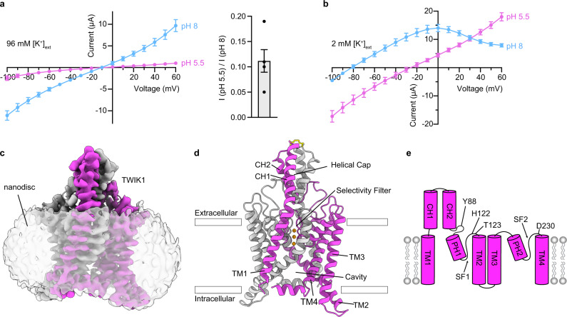
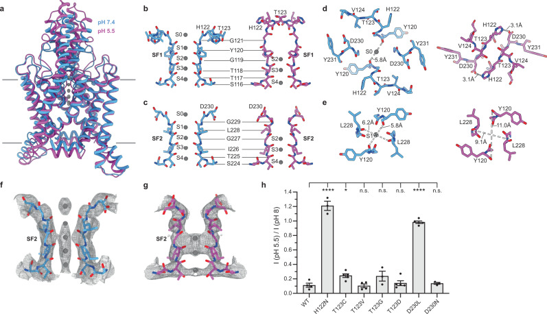
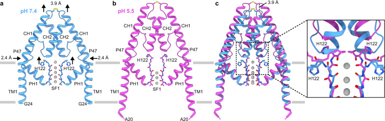
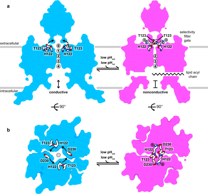
TWIK1 (K2P1.1, KCNK1) is a widely expressed pH-gated two-pore domain K+ channel (K2P) that contributes to cardiac rhythm generation and insulin release from pancreatic beta cells. TWIK1 displays unique properties among K2Ps including low basal activity and inhibition by extracellular protons through incompletely understood mechanisms. Here, we present cryo-EM structures of TWIK1 in lipid nanodiscs at high and low pH that reveal a previously undescribed gating mechanism at the K+ selectivity filter. At high pH, TWIK1 adopts an open conformation. At low pH, protonation of an extracellular histidine results in a cascade of conformational changes that close the channel by sealing the top of the selectivity filter, displacing the helical cap to block extracellular ion access pathways, and opening gaps for lipid block of the intracellular cavity. These data provide a mechanistic understanding for extracellular pH-gating of TWIK1 and illustrate how diverse mechanisms have evolved to gate the selectivity filter of K+ channels.


Conflict of interest statement

The authors declare no competing interests.

Figures

Fig. 1
Fig. 1. Structure and function of TWIK1.
a, b Current-voltage relationships from TWIK1-expressing cells at high and low pHext in (a) high [K+]ext and (b) low [K+]ext. a, ((right) Fraction of current remaining after extracellular acidification in high [K+]ext (IpH 5.5/IpH 8 at 60 mV = 0.11 ± 0.03, mean ± sem from n = 4 and 6 cells for high [K+]ext and low [K+]ext, respectively. c Cryo-EM map at pH 5.5 viewed from the membrane plane. Nanodisc is transparent and TWIK1 subunits are magenta and white. d TWIK1 model colored as in c with K+ ions orange and disulfide yellow. e Cartoon representation of a TWIK1 protomer with four transmembrane helices (TM1-TM4), two extracellular cap helices (CH1-CH2), two-pore helices (PH1–PH2), two selectivity filters (SF1–SF2), and key residues involved in extracellular pH-gating indicated. Source data for a, b are provided as a Source Data file.
Fig. 2
Fig. 2. TWIK1 conduction pathways at high and low pH.
a, b Structures of TWIK1 at (a) pH 7.4 and (b) pH 5.5 with the surface of conduction pathways shown in gray. c Radius of the central pore as a function of distance along the conduction pathway. Positions of the selectivity filter, lipid block at low pH, K+ coordination sites S0-S4, and T123 are indicated. d Radius of the extracellular ion pathways as a function of distance from the central conduction axis.
Fig. 3
Fig. 3. TWIK1 selectivity filter gate controlled by pHext.
a Overlay of open (pH 7.4, blue) and closed (pH 5.5, magenta) conformations of TWIK1 viewed from the membrane plane. be Comparisons of regions involved in the selectivity filter gate from open (left) and closed (right) TWIK1 structures. b SF1, c SF2, d the top of the selectivity filter, and e coordination site S1 are shown b, c from the membrane plane and d, e from the extracellular side. K+ coordination sites S0-S4 and intra-carbonyl or H122-D230 salt bridge distances are indicated. f, g Cryo-EM maps around the selectivity filter illustrating ion occupancy in f open and g closed structures. h Fraction of current remaining upon extracellular acidification (pHext = 5.5/pHext = 8.0 at 60 mV) for wild-type TWIK1 (0.11 ± 0.03) and mutants H122N (1.21 ± 0.06), T123C (0.24 ± 0.02), T123V (0.10 ± 0.02), T123G (0.24 ± 0.07), T123D (0.14 ± 0.03), D230L (0.98 ± 0.02), and D230N (0.14 ± 0.01). Mean plus s.e.m. were plotted for n = 4,3,5,4,3,5,5, and 3 cells, respectively. Differences were assessed with one-way analysis of variance (ANOVA) with Dunnett correction for multiple comparisons (*P = 0.0425; ****P < 0.0001; n.s. not significant). Source data for h are provided as a Source Data file.
Fig. 4
Fig. 4. Helical cap and transmembrane helix rearrangements in response to pHext.
ac View of TM1s, helical cap, PH1s, and SF1s at (a) pH 7.4, (b) pH 5.5, and (c) overlaid. Arrows highlight the upward movements of H122 and helical cap and the inward movement of TM1 in response to low pHext. c (inset) Zoomed in view of region boxed in c illustrating steric overlap between the selectivity filter in the low pHext structure and helical cap in the high pHext structure.
Fig. 5
Fig. 5. Structural model for pH-gating of the TWIK1 channel.
a, b Model for selectivity filter gating of TWIK1 gating by pHext viewed from (a) the membrane plane and (b) the extracellular side. TWIK1 is conductive at a high pH. At low pH, H122 protonation results in conformational changes that disrupt K+ coordination sites S0 and S1, seal the top of the selectivity filter, displace the helical cap to block extracellular ion access pathways, and dilate a lateral membrane opening to permit acyl chain binding to the channel cavity.

References

    1. Enyedi P, Czirják G. Molecular background of leak K+ currents: two-pore domain potassium channels. Physiol. Rev. 2010;90:559–605. doi: 10.1152/physrev.00029.2009. - DOI - PubMed
    1. Natale AM, Deal PE, Minor DL., Jr. Structural insights into the mechanisms and pharmacology of K2P potassium channels. J. Mol. Biol. 2021;433:166995. doi: 10.1016/j.jmb.2021.166995. - DOI - PMC - PubMed
    1. Lesage F, et al. TWIK‐1, a ubiquitous human weakly inward rectifying K+ channel with a novel structure. Embo J. 1996;15:1004–1011. doi: 10.1002/j.1460-2075.1996.tb00437.x. - DOI - PMC - PubMed
    1. Rajan S, Plant LD, Rabin ML, Butler MH, Goldstein SAN. Sumoylation silences the plasma membrane leak K+ channel K2P1. Cell. 2005;121:37–47. doi: 10.1016/j.cell.2005.01.019. - DOI - PubMed
    1. Chatelain FC, et al. TWIK1, a unique background channel with variable ion selectivity. Proc. Natl Acad. Sci. 2012;109:5499–5504. doi: 10.1073/pnas.1201132109. - DOI - PMC - PubMed

Publication types